Figures & data
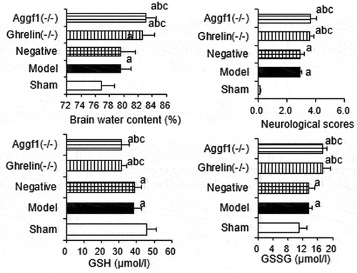
Table 1. siRNA primer information
Table 2. Evaluation standard for neurologic dysfunction
Table 3. Information about quantitative detection primers of target genes
Figure 1. Comparison of mRNA expression levels of target genes in rat brain tissues of each group after silence Ghrelin and Aggf1. (a showed Ghrelin mRNA expression levels, and b demonstrated Aggf1 mRNA expression levels. The comparison with sham group indicated aP < 0.05, the comparison with model group revealed bP < 0.05, and the comparison with negative group showed cP < 0.05).
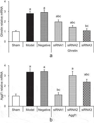
Figure 2. Comparison of scores of rat neurologic dysfunction in each group. (The comparison with sham group indicated aP < 0.05, the comparison with model group revealed that bP < 0.05, and the comparison with negative group showed that cP < 0.05).
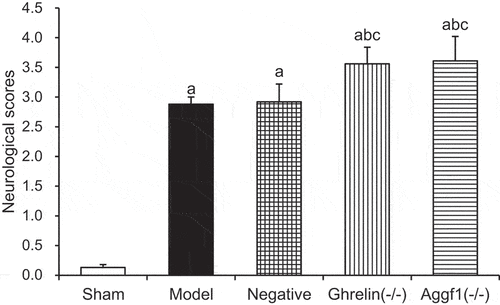
Figure 3. Comparison of rat cerebral water contents in each group. (The comparison with sham group indicated aP < 0.05, the comparison with model group showed bP < 0.05, and the comparison with negative group revealed that cP < 0.05).
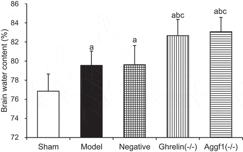
Figure 4. Comparison of Ghrelin and Aggf1 protein expression levels of rat brains in five groups (a showed Western blotting, b displayed Ghrelin protein expression levels, and c indicated Aggf1 protein expression levels. The comparison with sham group demonstrated P < 0.05, the comparison with model group revealed bP < 0.05, and the comparison with negative group suggested cP < 0.05).
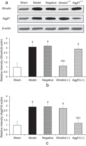
Figure 5. Comparison of p-Akt and caspase-3 expression levels of rat brains in five groups (a showed Western blotting, b displayed p-Akt protein expression levels, and c indicated caspase-3 protein expression levels. The comparison with sham group demonstrated P < 0.05, the comparison with model group revealed bP < 0.05, and the comparison with negative group suggested cP < 0.05).
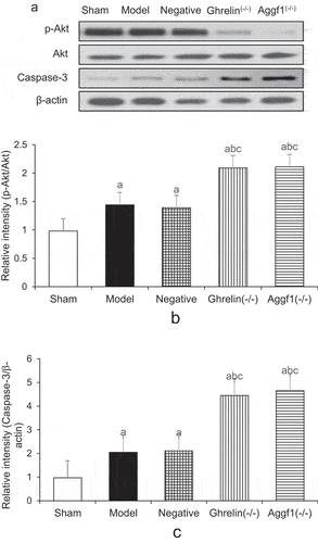
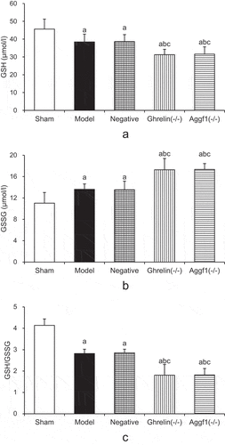
Figure 6. Comparison of GSH, GSSG, and GSH/GSSG levels of rat brains in each group. (a showed the comparison of GSH contents, b demonstrated the comparison of GSSG contents, and c presented GSH/GSGG ratio values. The comparison with sham group indicated aP < 0.05, the comparison with model group revealed bP < 0.05, and the comparison with negative group demonstrated cP < 0.05.).