Figures & data

Table 1. The relationship between LINC01133 expression and clinicopathological characteristics of PC patients
Figure 1. LINC01133 is upregulated in PC.
(a, b) LINC01133 expression in PAAD tissues and its correlation with the survival of PAAD patients from TCGA database. (c and d) LINC01133 expression was evaluated by RT-qPCR in tumor tissues and cell lines. (e) OS curves are illustrated based on the LINC01133 level. *P < 0.05; ** P < 0.01.
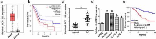
Figure 2. LINC01133 promotes PC malignancy.
(a) LINC01133 expression in PC cells after LINC01133 knockdown was evaluated by RT-qPCR. (b-f) Following LINC01133 depletion, cell proliferation was evaluated by CCK-8 (b), cell migration and invasion were detected by Transwell assays (C and D), cell apoptosis was assessed by TUNEL and flow cytometry assays (e and f). (g) The protein levels of apoptosis-related proteins (cleaved-Caspase-3, Bax, and Bcl-2) after silencing LINC01133 were detected by Western blot. (h and i) The impact of LINC01133 knockdown on xenograft tumor growth. (j) Change of Ki-67 level after LINC01133 depletion was evaluated by IHC. *** P < 0.001.
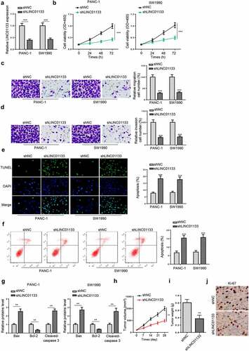
Figure 3. YY1 induces the upregulation of LINC01133 in PC cells.
(a) YY1 expression pattern in PC tissues from TCGA database. (b) YY1 binding motif and the prediction of YY1 binding sites within the promoter region of LINC01133 from JASPAR website. The binding sites between YY1 and LINC01133 promoter was verified in ChIP assay (c) and luciferase reporter assays (d). (e) Expression of LINC01133 in PANC-1 and SW1990 cells after depleting YY1. (f, g) YY1 expression in PC tissues was evaluated by RT-qPCR (F) and the correlation between expressions of YY1 and LINC01133 in tumor tissues was analyzed by Pearson’s analysis (g). (h) YY1 expression in PC tissues was detected by IHC *P < 0.05; ** P < 0.01.
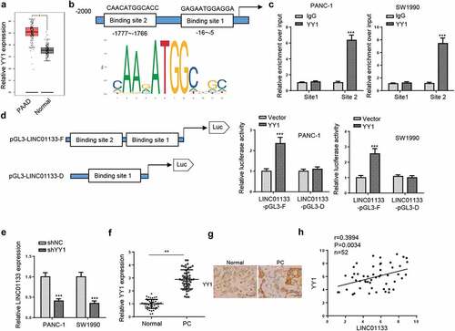
Table 2. The relationship between miR-199b-5p expression and clinicopathological characteristics of PC patients
Figure 4. LINC01133 functions as a ceRNA for miR-199b-5p.
(a) Subcellular localization of LINC01133 in PC cells. (b and c) Expression of miR-199b-5p in tumor and non-tumor tissues and cell lines was detected by RT-qPCR. (d) Starbase prediction of the binding sites between LINC01133 and miR-199b-5p. (e) Validation of the binding sites between LINC01133 and miR-199b-5p by luciferase reporter assay. (f) RT-qPCR evaluated the change of miR-199b-5p expression in PANC-1 and SW1990 cells following transfection with shLINC01133 or pcDNA3.1/LINC01133. (g) Correlation between levels of LINC01133 and miR-199b-5p in PC tissues. *P < 0.05; ** P < 0.01; *** P < 0.001.
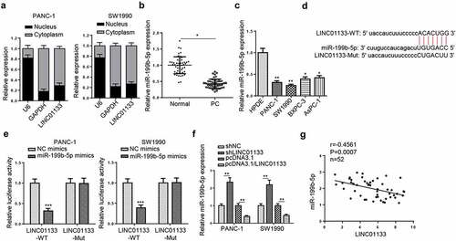
Figure 5. LINC01133 regulates PC progression through miR-199b-5p.
(a) RT-qPCR results of miR-199b-5p expression in PANC-1 and SW1990 cells following depletion of LINC01133 and miR-199b-5p. (b-e) Cell proliferation (b), cell migration (c), cell invasion (d) and cell apoptosis (e) following co-transfection of shLINC01133 with miR-199b-5p inhibitor were evaluated by CCK-8, Transwell and TUNEL assays. ** P < 0.01; *** P < 0.001.

Figure 6. LINC01133 regulates MYRF expression via miR-199b-5p.
(a) microT, miRanda, miRmap, PITA, and RNA22 databases were used to predict putative downstream genes for miR-199b-5p. (b) TCGA data analysis of MYRF expression in PAAD. (c) Predicted binding sites between miR-199b-5p and MYRF. (d) Analysis of MYRF luciferase activity in PC cells after transfection of miR-199b-5p mimics. (e and f) RT-qPCR and Western blot detected the levels of MYRF in PC cells following transfection of shLINC01133 or shLINC01133+ miR-199b-5p inhibitor. (g) Correlation between MYRF and miR-199b-5p expression in PC tissues. ** P < 0.01; *** P < 0.001.
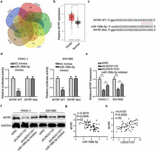
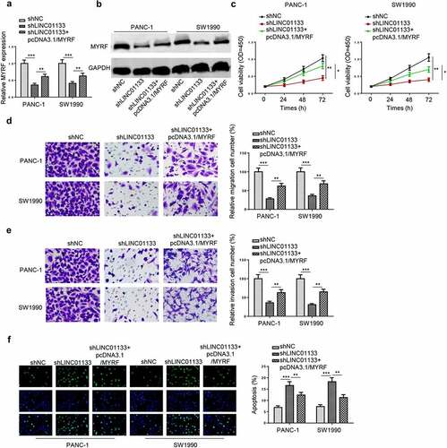
Figure 7. LINC01133 regulates PC malignancy in vitro by positively mediating MYRF.
(a and b) RT-qPCR and Western blot assessed the expression of LINC01133 in PC cells following transfection of shLINC01133 and shLINC01133+ pcDNA3.1/MYRF. (c-f) After designated cell treatment, cell proliferation (c), migration (d), invasion (e) and apoptosis (f) were evaluated respectively. ** P < 0.01; *** P < 0.001.