
Figures & data
Figure 1. LEMD1 expression is markedly increased in colon cancer tissues and cell lines and concerned with poor prognosis. (a) Differential LEMD1 expression in tumor and adjacent tissues, the correlation of LEMD1 expression with (b) pathological stages, (c) overall survival rate and (d) disease-free survival rate in colon cancer patients were analyzed by GEPIA database. *P < 0.05. (e) RT-qPCR and (f) western blot ascertained LEMD1 expression in colon cancer cell lines and normal intestinal epithelial HIEC cells. ***P < 0.001 vs. HIEC.
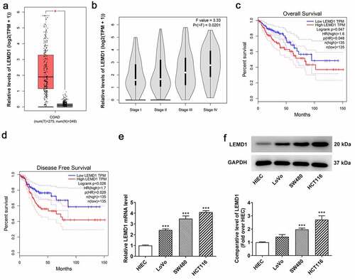
Figure 2. Insufficiency of LEMD1 hampers the proliferation of HCT116 cells. (a) RT-qPCR and (b) western blot analysis of the interference efficacy of LEMD1-targeted shRNAs. The proliferative ability of HCT116 cells was assessed by (c) CCK-8 and (d) colony formation assays. (e) The protein levels of Ki-67 and PCNA were determined by western blot. (f) Wound healing assay appraised cell migration. Magnification, x100. (g) Transwell assay estimated cell invasion. Magnification, x100. (h) MMP2 and MMP9 protein levels were determined by western blot. *P < 0.05, ***P < 0.001 vs. sh-NC.
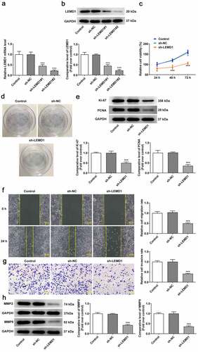
Figure 3. LEMD1 knockdown suppresses angiogenesis in colon cancer. (a) Tube formation assay detected angiogenesis. Magnification, x4. (b) The protein levels of VEGF and p-VEGFR2/VEGFR2 were determined by western blot. **P < 0.01, ***P < 0.001 vs. sh-NC.
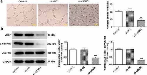
Figure 4. The up-regulation of LEMD1 in colon cancer is mediated by SOX4 transcription factor. (a) The potential binding sites between LEMD1 promoter and SOX4 were predicted by JASPAR database. (b) Differential SOX4 expression in tumor and adjacent tissues and (c) the correlation between LEMD1 and SOX4 were analyzed by GEPIA database. (d) RT-qPCR and (e) western blot ascertained SOX4 expression in HCT116 and HIEC cells. **P < 0.01, ***P < 0.001 vs. HIEC. (f) RT-qPCR and (g) western blot tested transfection efficiency of Ov-SOX4. ***P < 0.001 vs. Ov-NC. (h) Luciferase reporter assay verified the luciferase activity of WT and MUT LEMD1 promoter. ***P < 0.001 vs. LEMD1+ Ov-NC. (i) ChIP assay identified the accumulation of LEMD1 promoter in SOX4 antibody. ***P < 0.001 vs. IgG. (j) RT-qPCR and (k) western blot ascertained LEMD1 expression after SOX4 was overexpressed. **P < 0.01, ***P < 0.001 vs. Ov-NC.
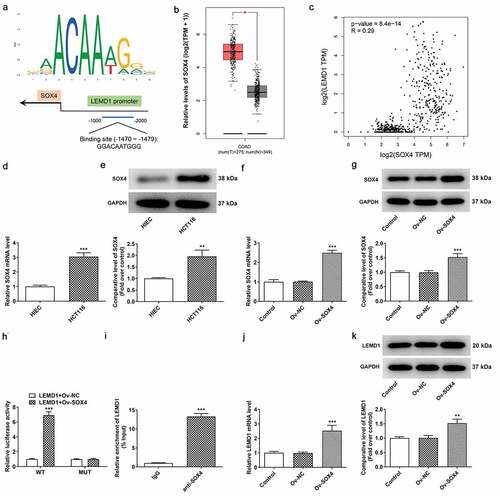
Figure 5. LEMD1 mediated by SOX4 contributes to cell proliferation, migration and invasion in colon cancer cells. The proliferative ability of HCT116 cells was assessed by (a) CCK-8 and (b) colony formation assays. (c) The protein levels of Ki-67 and PCNA were determined by western blot. (d) Wound healing assay appraised cell migration. Magnification, x100. (e) Transwell assay estimated cell invasion. Magnification, x100. (f) MMP2 and MMP9 protein levels were determined by western blot. *P < 0.05, ***P < 0.001 vs. control; ##P < 0.01, ###P < 0.001 vs. sh-LEMD1+ Ov-NC.
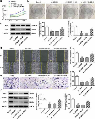
Figure 6. LEMD1 mediated by SOX4 induces angiogenesis in colon cancer cells. (a) Tube formation assay detected angiogenesis. Magnification, x4. (b) The protein levels of VEGF and p-VEGFR2/VEGFR2 were determined by western blot. ***P < 0.001 vs. control; ##P < 0.01, ###P < 0.001 vs. sh-LEMD1+ Ov-NC.
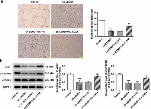
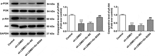
Figure 7. SOX4-induced LEMD1 activates the PI3K/AKT signaling in colon cancer cells. Western blot analyzed the protein levels of p-PI3K/PI3K and p-AKT/AKT in HCT116 cells. ***P < 0.001 vs. control; ###P < 0.001 vs. sh-LEMD1+ Ov-NC.
Data availability statement
All data included in this study are available upon request through contact with the corresponding author.