Figures & data
Figure 1. ARAP1-AS1 knockdown suppresses LUAD cell growth and metastasis. (a) GEPIA website revealed ARAP1-AS1 expression in normal tissues (n = 347) and LUAD tissues (n = 483). (b) ARAP1-AS1 expression in LUAD cell lines (A549 and H1975) and a normal lung epithelial cell line (BEAS-2B) were evaluated by RT-qPCR. (c) The interfering efficiency of ARAP1-AS1 in LUAD cells was subjected to RT-qPCR. (d-e) MTT and colony formation assays were performed to detect LUAD cell proliferation after ARAP1-AS1 downregulation. (f-g) LUAD cell invasion and migration after ARAP1-AS1 knockdown were respectively measured by Transwell invasion and wound healing assays. (*) p < 0.05, (**) p < 0.01, (***) p < 0.001.
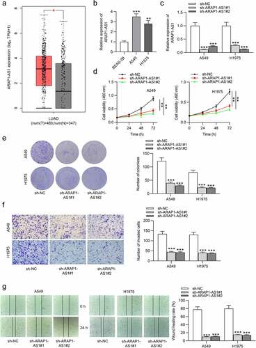
Figure 2. ARAP1-AS1 overexpression facilitates LUAD cell growth and metastasis. (a) The overexpression efficiency of ARAP1-AS1 in LUAD cells was detected by RT-qPCR. (b-c) MTT and colony formation assays were performed to measure LUAD cell proliferation after ARAP1-AS1 overexpression. (d-e) LUAD cell invasion and migration after overexpressing ARAP1-AS1 were respectively examined by Transwell invasion assay and wound healing assay. (*) p < 0.05, (**) p < 0.01, (***) p < 0.001.
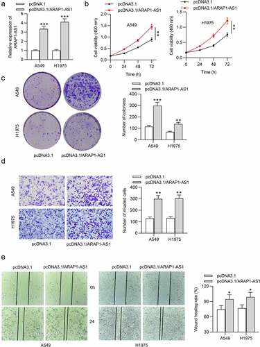
Figure 3. ARAP1-AS1 negatively modulates ARAP1 expression. (a) Subcellular fraction assay was performed for determining the cellular localization of ARAP1-AS1 in LUAD, with U6 and GAPDH as internal controls. (b-c) The influencing of ARAP1-AS1 silencing on mRNA and protein levels of its sense RNA ARAP1 were evaluated by RT-qPCR and western blotting. (d) The luciferase activity of plasmids containing ARAP1 promoter in LUAD cells after ARAP1-AS1 downregulation was evaluated by luciferase reporter assay. (e) ARAP1 expression in LUAD cells versus normal cells was assessed by RT-qPCR. (f) ARAP1 expression in normal tissues (n = 347) and LUAD tissues (n = 483) was shown at GEPIA website. (g) The correlation between ARAP1 expression level and poor prognosis was predicted according to Kaplan-Meier Plotter website. (*) p < 0.05, (**) p < 0.01, (***) p < 0.001.
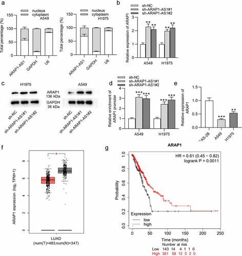
Figure 4. ARAP1-AS1 recruits EZH2 to regulate ARAP1 expression. (a) EZH2 expression in normal tissues (n = 59) and LUAD tissues (n = 526) was predicted at starBase website. (b) RT-qPCR was employed to detect EZH2 expression in LUAD cells versus normal cells. (c-d) The knockdown efficiency of EZH2 in LUAD cells was subjected to RT-qPCR and western blotting. (e-f) RT-qPCR and western blotting were carried out to detect the effects of EZH2 knockdown on ARAP1 expression and protein level. (g) A luciferase reporter assay was performed to measure the luciferase activity of plasmids containing ARAP1 promoter in LUAD cells after EZH2 knockdown. (h-i) The influence of EZH2 overexpression on ARAP1 expression in sh-ARAP1-AS1 transfected LUAD cells. (j) RIP assay was applied to evaluate the enrichment of ARAP1 promoter in H3K27 group in LUAD cells transfected with sh-ARAP1-AS1#1 and pcDNA3.1/EZH2. (*) p < 0.05, (**) p < 0.01, (***) p < 0.001.
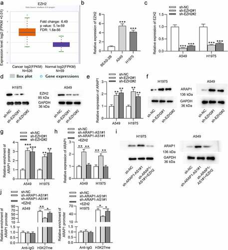
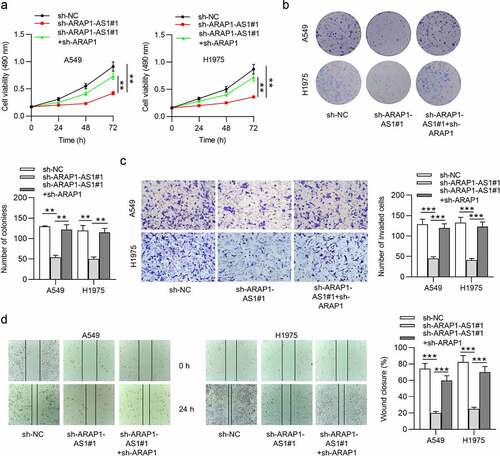
Figure 5. ARAP1 knockdown reverses the inhibition of ARAP1-AS1 on LUAD cell malignant behaviors. (a-b) MTT and colony formation assays were conducted to measure LUAD cell proliferation in sh-NC, sh- ARAP1-AS1#1, sh-ARAP1-AS1#1 + sh-ARAP1 groups. (c-d) Transwell invasion and wound healing assays were performed to evaluate LUAD cell invasion and migration in the above three groups. (*) p < 0.05, (**) p < 0.01, (***) p < 0.001.