Figures & data
Figure 1. Asiaticoside alleviated renal function in IRI mice. BUN (a) and Scr (b) in renal ischemia-reperfusion injury mice, followed by saline or asiaticoside injection at 0, day1, day3, or day7. **p < 0.01 vs IRI groups, ##p < 0.01 vs Sham group. (n = 5 in each group).
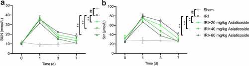
Figure 2. Asiaticoside inhibited the inflammation in renal ischemia-reperfusion injury mice. The levels of IL-1β (a), IL-6 (b), TNF-α (c), and IL-10 (d) in Sham group, IRI group, IRI with a low dose group (20 mg/kg), a medium dose group (40 mg/kg) and a high dose group (60 mg/kg) groups. **p < 0.01 vs IRI groups, ##p < 0.01 vs Sham group. (n = 5 in each group).
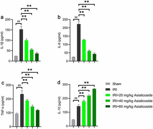
Figure 3. Asiaticoside reduced the infiltration of macrophages in renal IRI mice. The fluorescence intensity of F4/80 in Sham group, IRI group, IRI with a low dose group (20 mg/kg), a medium dose group (40 mg/kg) and a high dose group (60 mg/kg) groups.
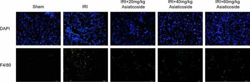
Figure 4. Asiaticoside attenuates LPS-induced inflammation in Raw264.7 cells. The content of IL-1β (a), IL-6 (b), TNF-α (c), and IL-10 (d) in the culture medium of Raw264.7 cells of different groups (control group, LPS group and LPS with different doses of asiaticoside). **p < 0.01 vs LPS groups, ##p < 0.01 vs control group.
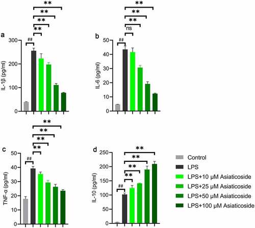
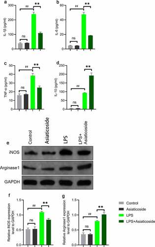
Figure 5. Asiaticoside improved LPS-induced inflammation and transformed M1 and M2 marker expressions in Raw264.7 cells. The levels of IL-1β (a), IL-6 (b), TNF-α (c), and IL-10 (d) in the culture medium of Raw264.7 cells of different groups (control group, asiaticoside (100 μM) group, LPS group and LPS+ asiaticoside (100 μM) group). (e)After immunoblotting, the levels of iNOS and Arginase1 were identified using their specific antibodies. The expression s of iNOS (f) and Arginase1 (g) are demonstrated. **p < 0.01 vs LPS group, ##p < 0.01 vs control group, nsp > 0.05 vs control group.