Figures & data
Figure 1. HSF2BP expression and prognosis in LUAD based on TCGA datasets. (a) The mRNA expression in LUAD and normal samples. (b) The expression in LUAD and paired normal samples. (c) Correlation analysis between HSF2BP expression and tumor stage. (d) Survival curves of HSF2BP in LUAD. (e) Cox regression analysis of HSF2BP and clinical parameters.
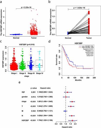
Figure 2. The expression of HSF2BP in LUAD tissues and cells. (a-b) The mRNA expression of HSF2BP in LUAD and normal tissues. (c) The protein expression of HSF2BP in LUAD and adjacent tissues which was detected by western blot. (d) The protein expression of HSF2BP in LUAD and adjacent tissues which was detected by IHC. (e) The protein expression of HSF2BP in LUAD cell lines and BEAS-2B. Note: * p < 0.05, ** p < 0.01, *** p < 0.001.
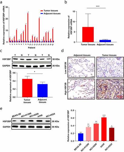
Figure 3. HSF2BP promotes the proliferation and migration of LUAD cells. (a) The transfection efficacy of lentivirus down-regulating HSF2BP in A549 and NCI-H209 cells. (b) The cell viability of A549 and NCI-H209 cells were determined by CCK-8 assay. (c) The colony formation assay was used to measure proliferation in LUAD cells. (d) Wound healing assay in A549 and NCI-H209 cells. (e) Transwell assay was used to determine the migration ability of LUAD cells. Note: * p > 0.05, ** p < 0.01, *** p < 0.001.
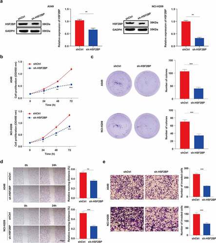
Figure 4. HSF2BP is involved in the regulation of MAPK signaling pathway. (a) GSEA analysis of HSF2BP. (b) The expression level of p-ERK, total ERK, p-JNK, total JNK, p-p38 and total p38 in LUAD cells by western blot.
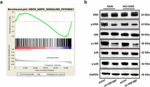
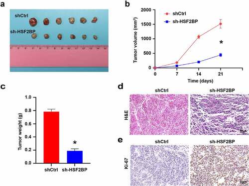
Figure 5. HSF2BP promotes the tumor growth in vivo. (a) The tumor formed by shCtrl and sh-HSF2BP cells. (b) Tumor volume. (c) Tumor weight. (d) HE staining. (e) Ki67 expression detected by immunohistochemistry. Note: * p < 0.001.