
Figures & data
Table 1. The sequences of all primers used in qRT-PCR
Figure 1. Effects of TWEAK and NF-κB on astrocytes. (a) Relative expressions of mRNA-TWEAK and mRNA-NF-κB were confirmed by qRT-PCR assays after the interference of TWEAK or NF-κB in astrocytes. (b) CCK-8 assay analysis of astrocyte viability after the interference of TWEAK or NF-κB. (c) Relative expressions of TNF-α and IL-1β were determined by ELISA after the interference of TWEAK or NF-κB. All the data were represented as mean ± SD, *p < 0.05 and **p < 0.05.
Figure 2. Interaction mechanisms of TWEAK and NF-κB. (a) Relative expression of mRNA-NF-κB and mRNA-TWEAK were confirmed by qRT-PCR assays after the interference of TWEAK or NF-κB in astrocytes. (b) Levels of p50 were detected by western blot analysis. Lamin B was used as the internal reference. (c) Expression of mRNA-TWEAK was confirmed by PCR assay in CHIP. pc: pcDNA3.1 empty vector. pc-NF-κB: pcDNA3.1/NF-κB. All the data were represented as mean ± SD, **p < 0.05 and ***p < 0.001.
Figure 3. Effects of TWEAK and NF-κB on autophagy. (a) Electron micrograph of autophagic cells. (b) Levels of LC3 and Beclin-1 were detected by western blot analysis after the interference of TWEAK or NF-κB. β-actin was used as the internal reference. (c) IF images of LC3 in astrocyte after the interference of TWEAK and NF-κB. All the data were represented as mean ± SD, **p < 0.05.
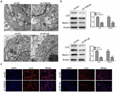
Figure 4. Autophagic cell death participated in the effect of TWEAK/NF-κB on astrocytes. (a) Electron micrograph of the lysosome. (b) Expression levels of Bax, Bcl-2 and Beclin-1 were determined by western blot after the interference of TWEAK or NF-κB. β-actin was used as the internal reference. (c) CCK-8 assay analysis of astrocyte viability after the interference of TWEAK or NF-κB and adding Rap. (d) Relative expressions of IL-1β and TNF-α in astrocytes were determined by ELISA after the interference of NF-κB or TWEAK and adding Rap. All the data were represented as mean ± SD, **p < 0.05, ***p < 0.001, #p < 0.05 and ##p < 0.01.
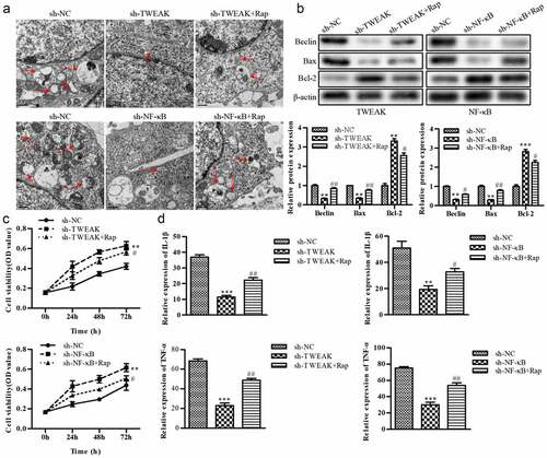
Figure 5. Effect of TWEAK/NF-κB on co-cultured neurons. (a) The relative expression values of TNF-α and IL-1β in the co-cultured neurons were determined by ELISA after the interference of TWEAK or NF-κB. (b) The expression level of GAP-43 was determined by western blot after the interference of TWEAK or NF-κB. β-actin was used as the internal reference. (c) IF images of GAP-43 in the co-cultured neurons after the interference of TWEAK or NF-κB. All the data were expressed as the means ± SD. *p < 0.05 and **p < 0.05.
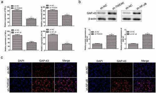
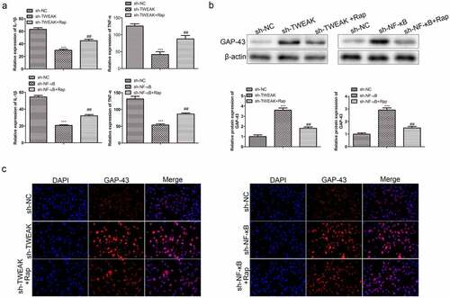
Figure 6. Autophagic cell death was involved in the effect of TWEAK/NF-κB on astrocytes co-cultured neurons. (a) Relative expressions of TNF-α and IL-1β in the co-cultured neurons were determined by ELISA after the interference of TWEAK/NF-κB and adding Rap. (b) Expression levels of GAP-43 in the co-cultured neurons were determined by western blot after the interference of TWEAK or NF-κB and adding Rap. β-actin was used as the internal reference. (c) IF images of GAP-43 in the co-cultured neurons after the interference of TWEAK or NF-κB and adding Rap. All the data were expressed as the means ± SD. ***p < 0.001 and ##p < 0.01.
Supplemental Material
Download Zip (143.6 MB)Data availability statement
All data generated or analysed during this study are included in this published article.