Figures & data
Table 1. Basic clinical data of participants
Figure 1. The expression of miR-101-3p in PARDS patients. A. The low expression of miR-101-3p in children with PARDS. B. The expression of miR-101-3p in the mild, moderate, and severe PARDS patients. ***P < 0.001, compared with healthy individuals, ###P < 0.001, compared with mild group.
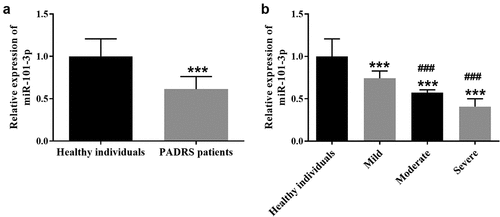
Table 2. Multiple linear regression analysis on variables associated with miR-101-3p
Figure 2. The AUC of miR-101-3p was 0.939 with a sensitivity of 90.2% and a specificity of 85.7%.
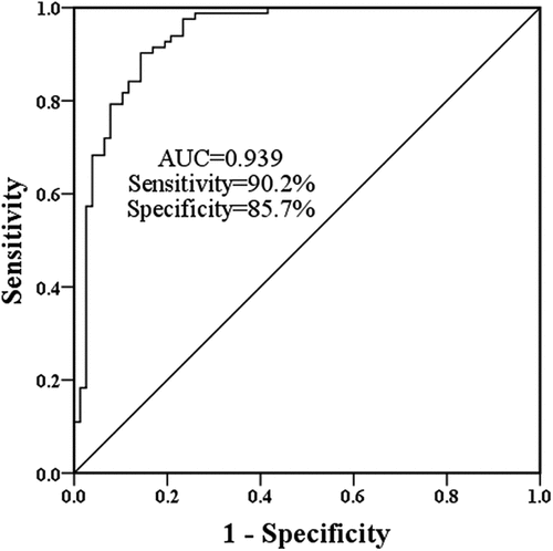
Figure 3. The role of miR-101-3p in LPS-evoked cell models. A. LPS inhibited miR-101-3p expression, but transfection altered miR-101-3p expression. B The findings of CCK-8 on A549 cells showed overexpression of miR-101-3p restricted the influence of LPS and interference of miR-101-3p accelerated the effects of LPS. C The beneficial effects of miR-101-3p on cell apoptosis. D The inhibitory effect of miR-101-3p on the inflammatory biomolecules in A549 cells. ***P < 0.001, compared with the control cells, #P < 0.05, ##P < 0.01, ###P < 0.001, compared with the LPS group.
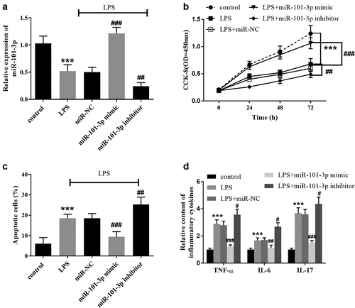
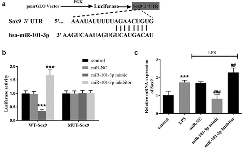
Figure 4. Sox9 is a target gene of miR-101-3p. A Visualization of the binding sites between Sox9 and miR-101-3p. B The luciferase activity was decreased in the miR-101-3p mimic subgroup and increased in the miR-101-3p inhibitor subgroup in WT-Sox9 group. C Sox9 mRNA expression was increased in the LPS group, which was compounded by the shift in miR-101-3p expression in A549 cell models. ***P < 0.001, compared with the control cells, ##P < 0.01, ###P < 0.001, compared with the LPS group.