
Figures & data
Figure 1. The protective effect of EE on cerebral I/R injury and apoptosis of hippocampal neuron cells. (a) Effect of EE on infarct volume of rat brain subjected to I/R injury. (b) Infarction ratios from (A) were presented as mean ± SEM. (c) Light microscopy photomicrographs depicting sections from brain of rats receiving sham operation, I/R, or I/R plus EE. (d) Light microscopy photomicrographs depicting sections from hippocampus of rats receiving sham operation, I/R, or I/R plus EE. (e) Apoptosis analysis of hippocampal neuron cells in rats treated with sham operation, I/R, or I/R plus EE. (f) The apoptosis rate from (E). (g) CCK-8 assay of hippocampal neuron cells in rats treated with sham operation, I/R, or I/R plus EE. Error bars ± SEM, *p < 0.05.
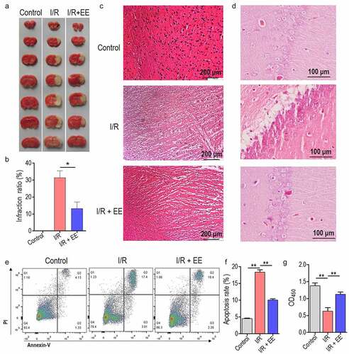
Figure 2. The apoptosis in I/R-injured brain and hippocampus tissues and the expressions of caspase 3, 6, and 7 in I/R-treated hippocampal neuron cells could be relieved by EE administration. (a) Representative images of TUNEL staining for coronary brain tissues receiving sham operation, I/R, or I/R plus EE. (b) Representative images of TUNEL staining for hippocampus tissue receiving sham operation, I/R, or I/R plus EE. The expression analysis of caspase 3, 6, and 7 both in mRNA (c) and protein (d) levels in hippocampal neuron cells of I/R-treated rats in the absence and presence of EE by real time PCR and western blotting. (e) The relative density of protein level of caspase 3, 6, and 7 from (D). Data were presented as mean ± SEM, **p < 0.01.
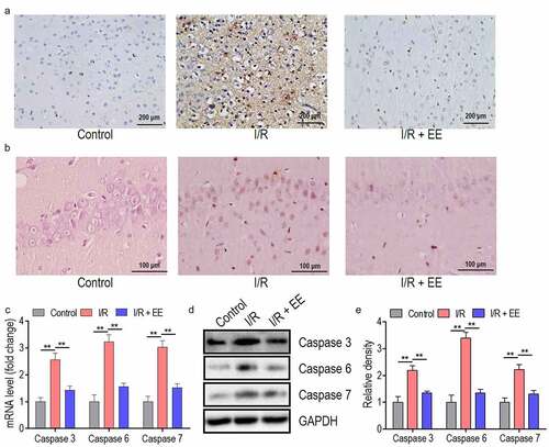
Figure 3. EE has an anti-oxidative effect on I/R-treated hippocampal neuron cells. EE administration reduced the ROS production (a) and MDA content (b) and increased the activities of SOD (c) and GSH peroxidase (d) on I/R-induced hippocampal neuron cells. Data were presented as mean ± SEM, **p < 0.01.
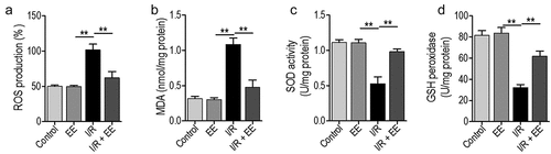
Figure 4. EE-treated hippocampal neuron cells show enhanced expression of Htr2c. (a) Unsupervised hierarchical clustering of thirty-eight vital genes in hippocampal neuron cells of I/R-treated rats in the absence and presence of EE. (b) The mRNA expression of Htr2c in the presence of EE. (c) The protein expression of Htr2c in the presence of EE. (d) The relative density of protein level of Htr2c from (C). Data were presented as mean ± SEM, **p < 0.01.
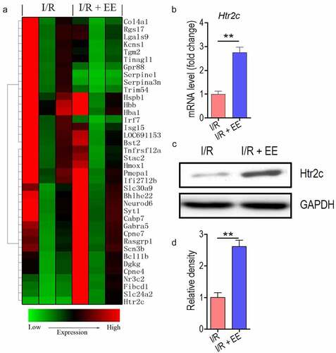
Figure 5. Htr2c contributes to anti-apoptosis effect of EE on I/R-treated hippocampal neuron cells. (a and b) The expression analysis of Htr2c both in mRNA (a) and protein (b) levels in hippocampal neuron cells treated with siRNA-Htr2c-1 to 3 by real time PCR and western blotting. (c) The relative density of protein level of Htr2c from (B). (d) The apoptosis analysis of hippocampal neuron cells of I/R rats treated simultaneously without or with EE and EE plus siRNA-Htr2c-3. (e) The apoptosis rate from (D). Data were presented as mean ± SEM.
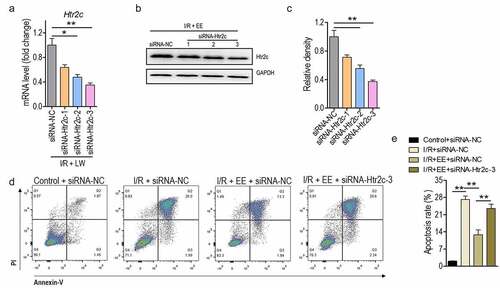
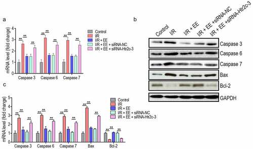
Figure 6. EE inhibits the expressions of caspase 3, 6, and 7 through Htr2c. (a) The expression analysis of caspase-3, −6 and −7 in mRNA levels in hippocampal neuron cells of I/R rats treated simultaneously without or with EE, EE plus siRNA-NC, and EE plus siRNA-Htr2c-3. (b) Western blotting of caspase-3, −6 and −7, Bax, and Bcl2 in hippocampal neuron cells of I/R rats treated simultaneously without or with EE, EE plus siRNA-NC, and EE plus siRNA-Htr2c-3. (c) The relative density of protein level of caspase 3, caspase 6, caspase 7, Bax, and Bcl-2 from (B).Data were presented as mean ± SEM, *p < 0.05, **p < 0.01.
Supplemental Material
Download Zip (1.5 MB)Data Availability Statement
Data available on request.