Figures & data
Table 1. The primer sequences of RIG-1, IFN-β and GAPDH for RT-qPCR
Figure 1. Isolation of exosomes from TAFs cells.(a) Flow cytometry was used to detect the expression of α-SMA, CD34, and CD45. (b) Representative TEM images of exosomes derived from TAFs cells showing morphology and size range. Scale bar = 200 nm. (c) Representative Western blot showing the expression of exosome markers (CD63, CD9, and CD81).
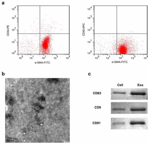
Figure 2. Exosomes induce proliferation and inhibit apoptosis of ESCC cells.(a) MTT assay to examine the cell proliferation. The data were shown as the optical density measured at 490 nm. (b) Flow cytometry was applied to assess cell apoptosis. (c) Western blot of Bcl-2, Bax and caspase-3. *, p < 0.05; **, p < 0.01.
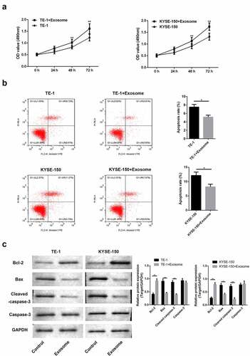
Figure 3. TAFs derived exosomes affect the chemosensitivity of ESCC cells to cisplatin via RIG-I/IFN-β signaling.(a) RT-PCR analysis of RIG-I/IFN-β. (b) Western blot analysis of RIG-I/IFN-β. **, p < 0.01; ***, p < 0.001.
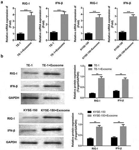
Figure 4. TAFs derived exosomes affect the chemosensitivity of ESCC cells to cisplatin.(a) MTT assay was employed to examine the cell proliferation. (b) Flow cytometry was applied to analyze cell apoptosis. *, p < 0.05; **, p < 0.01.
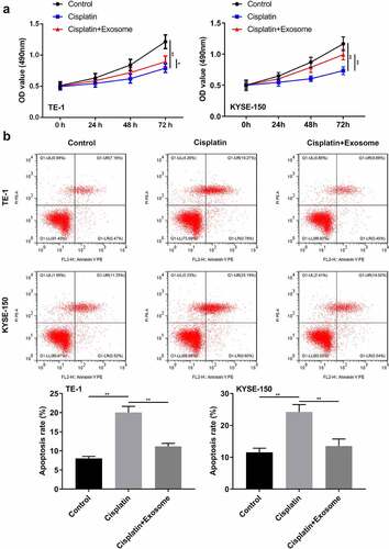
Figure 5. TAFs derived exosomes affect the chemosensitivity of ESCC cells to cisplatin via RIG-I/IFN-β signaling.(a) RT-PCR analysis of RIG-I/IFN-β. (b) Flow cytometry was applied to assess cell apoptosis. *, p < 0.05; **, p < 0.01.
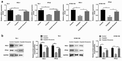
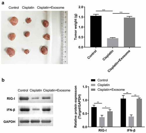
Figure 6. TAFs derived exosomes affect the cisplatin chemosensitivity in vivo.(a) Images and tumor weight of the xenograft tumors. (b) Western blot analysis of RIG-I/IFN-β. *, p < 0.05; **, p < 0.01.