Figures & data
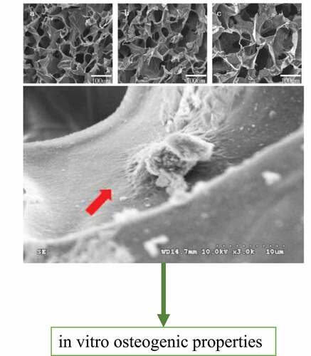
Table 1. RT–qPCR target gene primer sequences.
Figure 1. TEM observations of the pearl powder after grinding.
Figure 2. Morphology of the composite artificial bone. A. Image of the nanopearl powder/C-HA/rhBMP-2 artificial bone as viewed with the naked eye. B. SEM image of a cross-section of the nanopearl powder/C-HA/rhBMP-2 composite artificial bone at 100X magnification.
Figure 3. XRD (X-ray diffraction) spectrum of the nanopearl powder/C-HA/rhBMP-2 composite artificial bone.
Figure 4. Curve showing the sustained release of rhBMP-2 from the nanopearl powder/C-HA/rhBMP-2 composite artificial bone.
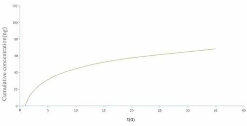
Figure 5. Adhesion and growth of MC3T3-E1 cells on the surface of the artificial bone. Mouse MC3T3-E1 cells were cocultured with the nanopearl powder/C-HA/rhBMP-2 composite artificial bone, and the morphology of the cells in the three-dimensional artificial bone was observed using SEM.
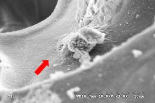
Figure 6. CCK-8 assays of cell proliferation. MC3T3-E1 cells were cocultured with sterilized composite artificial bone materials (C-HA composite artificial bone (S1), nanopearl powder/C-HA composite artificial bone (S2), and nanopearl powder/C-HA/rhBMP-2 composite artificial bone (S3)).
Figure 7. ALP activity in MC3T3-E1 cells from each group increased (* indicates P < 0.05) after growth on C-HA composite artificial bone (S1), nanopearl powder/C-HA composite artificial bone (S2), and nanopearl powder/C-HA/rhBMP-2 composite artificial bone (S3).
Figure 8. Expression of the ColαI, OCN, OPN and Runx2 genes (* indicates P < 0.05). MC3T3-E1 cells were cocultured with sterilized composite artificial bone materials (C-HA composite artificial bone (S1), nanopearl powder/C-HA composite artificial bone (S2), and nanopearl powder/C-HA/rhBMP-2 composite artificial bone (S3)). A. RT–qPCR analysis of the expression of the ColαI, OCN, OPN and Runx2 mRNAs. B. Western blot analysis of the expression of the ColαI, OCN, OPN and Runx2 proteins.