Figures & data
Figure 1. METTL3 and METTL14-mediated m6A modifications sustained high-levels of miR-380-3p in PC tissues and cells. (a) The expression levels of miR-380-3p in the PC patients’ normal and cancer tissues were analyzed by Real-Time qPCR. (b) PC patients’ prognosis was analyzed by performing the Kaplan-Meier survival analysis. (c) The expression status of miR-380-3p in the normal HPDE cells and PC cells were respectively determined. The silencing vectors for (d) METTL3 and (e) METTL14 were delivered into the PC cells, and the vectors transfection efficiency was examined by conducting Real-Time qPCR. (f, g) The effects of METTL3 and METTL14 knockdown on miR-380-3p levels in the PC cells were examined. Individual experiment was repeated for at least 3 times, and *P < 0.05 was deemed as statistical significance.
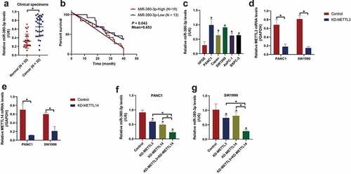
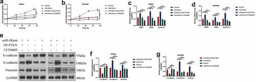
Figure 2. MiR-380-3p was verified as an oncogene to promote cell proliferation, EMT and tumorigenesis in PC cells. (a, b) CCK-8 assay revealed that miR-380-3p promoted cell proliferation in the PANC1 and SW1990 cells, which was dependent on culturing time. (c, d) The expression levels of cell-cycle associated genes, including CDK2, CDK6, and Cyclin D1, were determined by Real-Time qPCR. (e-g) Western Blot analysis confirmed that miR-380-3p downregulated E-cadherin, whereas upregulated N-cadherin and Vimentin to accelerate EMT process in the PC cells. (h-j) The xenograft tumor-bearing mice models were established by using the PANC1 cells, and the results suggested that miR-380-3p promoted tumorigenesis of this PC cell line in vivo. (i) Ki67 expression levels were examined by immunohistochemistry staining assay. Individual experiment was repeated for at least 3 times, and *P < 0.05 was deemed as statistical significance.
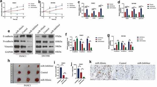
Figure 3. PTEN was predicted and validated as the downstream target of miR-380-3p. (a) The binding sites of miR-380-3p with the 3ʹUTR of PTEN mRNA were predicted by using the online starBase software. (b, c) The targeting sites between these two genes were verified by performing the dual-luciferase reporter gene system assay. (d) Real-Time qPCR and (e, f) Western Blot analysis were conducted to examine the mRNA and protein levels of PTEN in the PC cells. Individual experiment was repeated for at least 3 times, and *P < 0.05 was deemed as statistical significance.
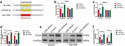
Figure 4. The Akt pathway was activated by miR-380-3p overexpression in a PTEN-dependent manner. The expression levels of Akt and p-Akt were determined by conducting Western Blot analysis in the (a) PANC1 and (b) SW1990 cells. Individual experiment was repeated for at least 3 times, and *P < 0.05 was deemed as statistical significance.
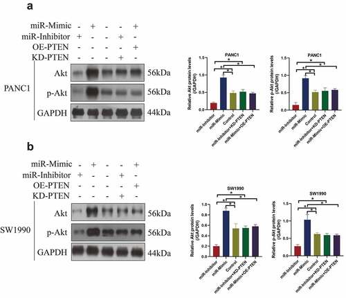
Figure 5. MiR-380-3p accelerated cancer malignancy in PC through modulating the PTEN-Akt signal pathway. (a, b) Cell proliferation abilities in the PANC1 and SW1990 cells were determined by performing the CCK-8 assay. (c, d) The Real-Time qPCR analysis was used to detect the mRNA levels of CDK2, CDK6, and Cyclin D1 in the PC cells. (e-g) The expression levels of the EMT-associated biomarkers were examined by performing the Western Blot analysis. Individual experiment was repeated for at least 3 times, and *P < 0.05 was deemed as statistical significance.