
Figures & data
Figure 1. Schematic representation of β-catenin protein structure.
Notes: The diagram encapsulates the principal structural breakdown of β-catenin where the three domains – N terminal domain (amino acids 1-150), armadillo domain with armadillo repeat R1 to R12 (amino acids 141-664), and C terminal domain (amino acids 665-781) along with the major binding regions for E-cadherin, TCF, BCL9, APC/AXIN, and CBP/P300 in the respective domains are marked. The main phosphorylated residues (denoted using symbol “ ”) and most frequently mutated residues in various cancers (denoted using symbol “
 ”) are also included in the corresponding domains. Created with BioRender.com
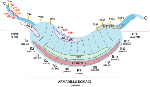
Figure 2. Mechanisms controlling β-catenin activation and dynamic localization.
Notes: ) schematically illustrates the regulation of β-catenin stability and localization by the canonical Wnt signaling pathway. In the Wnt ‘OFF’ state, the WNT ligands are absent and consequently, the entry of β-catenin into the nucleus is limited by the proteasomal degradation of β-catenin in the cytoplasm, which is facilitated by the phosphorylation of the protein by the AXIN-APC1-GSK3β-CK1 Destruction Complex (DC). Upon Activation, WNT ‘ON,’ the binding of WNT ligands to their frizzled receptor complex results in the recruitment of Disheveled and activates a series of events leading to disassembly of the destruction complex. This leads to cytoplasmic accumulation and subsequent translocation of β-catenin into the nucleus where it serves its transcriptional purpose. represents the WNT independent modes of activation of β-catenin that promotes its nuclear localization. It includes activating phosphorylation moderated by receptor tyrosine kinases like EGFR, FGFR, and IGFR. Competition of PTEN with β-catenin for CAV1 and competition of Plakoglobin (γ-catenin) for E-cadherin are also mechanisms that lead to the upregulation of β catenin in the cytoplasm, and subsequent nuclear translocation. Created with BioRender.com
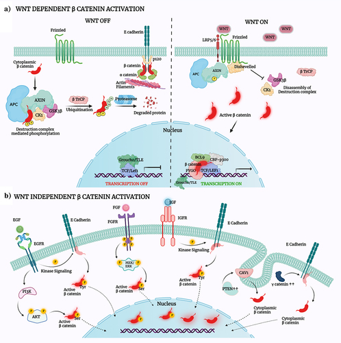
Figure 3. Transcriptional targets of β-catenin and their implications in promoting the hallmarks of cancer.
Notes: The figure encompasses a short list of genes that are under direct control of the β-catenin transcriptional activity which are broadly segregated based on their roles in promoting cancer progression and metastasis. It is noteworthy that the genes which are under transcriptional control of β-catenin have key roles to play in driving multiple hallmarks (i.e. replicative immortality and survival; angiogenesis; invasion and metastasis; metabolic reprogramming; chemoresistance; and immune invasion) of cancer making β-catenin a valuable therapeutic target. Created with BioRender.com
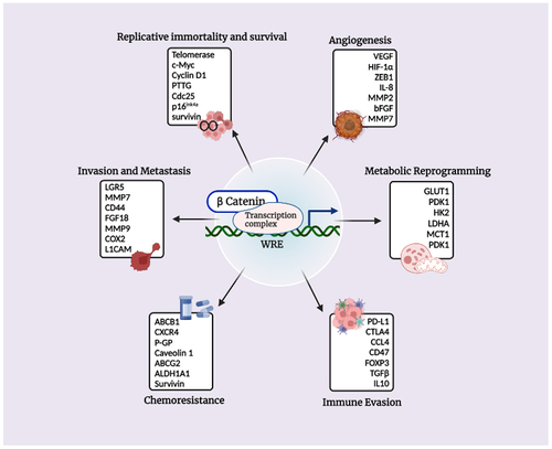
Figure 4. β-catenin enhanceosome complex and therapeutic alternatives.
Notes: A schematic illustration of the β-catenin transcriptional complex or the ‘enhanceosome.’ The inhibitors that target β-catenin and perturbs its interaction with BCL9, TCF, and CBP are included in respective boxes. A detailed list of β-catenin inhibitors and their mode of action is provided in . Created with BioRender.com
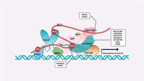
Table 1. A comprehensive list of β-catenin inhibitors with structures and mode of action.
Table 2. A comprehensive list of β-catenin natural inhibitors along with the structure and mode of action.
Data availability statement
Data sharing is not applicable to this article as no new data were created or analyzed in this study.