
Figures & data
Table 1. Preoperative laboratory examination of the patients.
Figure 1. The morphology of HCC-ZJ cells (a); immunohistochemistry for CK7 and CK19 (b); the karyotype of HCC-ZJ cells (c); the CK7 and CK19 positive rate of HCC-ZJ and intrahepatic cholangiocarcinoma (ICC) samples (n = 3) (d). * represents P < 0.05 and ** represents P < 0.01.
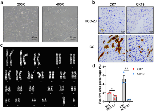
Table 2. Short tandem repeat (STR) identification of HCC-ZJ.
Figure 2. Statistics of the whole-exome sequencing. (a) the numbers and mutation types of genes (upper) and sites (lower) from INDELs; (b) the numbers and mutation types of genes (upper) and sites (lower) from SNPs; (c) the numbers of INDEL sites containing the corresponding number of bases; (d) the number of transitions and transversions based on the four kinds of bases.
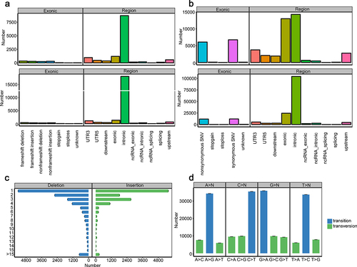
Figure 3. Circos plot describing the distribution of SNPs, INDELs, copy number variants (CNVs), and structural variations (SVs) in genome.
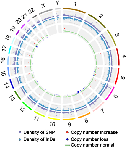
Table 3. Nonsynonymous single nucleotide variant (SNV) of HCC-ZJ cell by whole-exome sequencing.
Table 4. Frameshift insertion/deletion (INDEL) of HCC-ZJ cell by whole-exome sequencing.
Figure 4. In vitro characteristics of HCC-ZJ cells. (a) the proliferative potential of HCC-ZJ, Huh1, and HCCLM3 cells (n = 4); (b) colony formation ability of HCC-ZJ, Huh1, and HCCLM3 cells (n = 3); (c) migration and invasion abilities of HCC-ZJ, Huh1, and HCCLM3 cells (n = 3);(d) the sensitivity of HCC-ZJ, Huh1, and HCCLM3 to sorafenib (left) and lenvatinib (right) (n = 5).
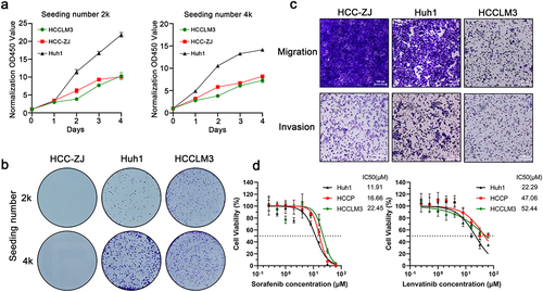
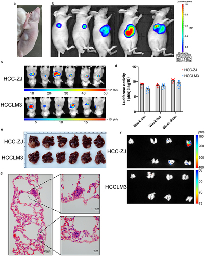
Figure 5. In vivo oncogenicity of the HCC-ZJ cell line. (a) subcutaneous inoculation of 1 × 10⁶ HCC-ZJ cells; (b) subcutaneous inoculation of 1 × 10⁶ HCC-ZJ-Luc cells; the luminescence reached 1 × 10⁸ (n = 5); (c) orthotopic tumorigenesis of 1 × 10⁶ HCC-ZJ-Luc and HCCLM3-Luc cells (n = 6) in the third week; (d) luciferase activity of HCC-ZJ-Luc and HCCLM3-Luc at the indicated time point; (e) dissection of orthotopic tumors (n = 6) in the third week; (f) imaging of newly dissected lungs from HCC-ZJ-bearing and HCCLM3-bearing mice showed that orthotopic tumors of HCC-ZJ could migrate into the lung (n = 6); the first lung of each group derived from healthy mice without orthotopic inoculation of HCC cells; (g) H&E staining demonstrating the lung metastasis of orthotopic HCC-ZJ.
Supplemental Material
Download Zip (55.9 MB)Data availability statement
The data presented in this study are available on request from the corresponding author. The data are not publicly available due to the patient’s privacy.