
Figures & data
Figure 1. Iron labeling by DAB-enhanced Perls' staining in wildtype and Hfe−/−xTfr2mut mouse brain at 3 months of age. Iron is mainly accumulated in myelinated structures and associated cells. Myelin (M), oligodendrocyte (O), blood vessel (V) and black arrows show neurons (N) which do not accumulate appreciable iron even in the Hfe−/−xTfr2mut mouse brain.
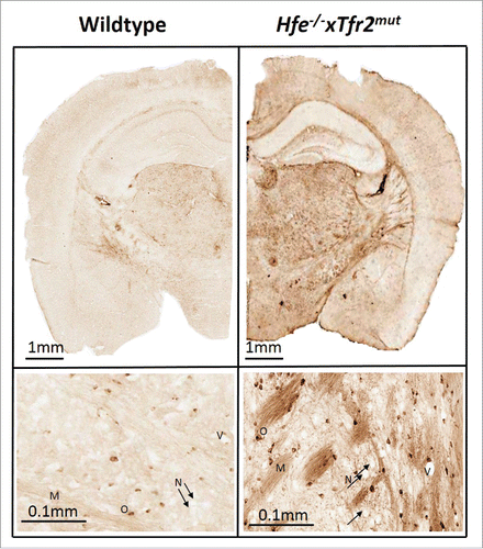
Table 1. Myelin-related genes identified as differentially expressed in i. the Hfe−/−xTfr2mut mice in comparison with wildtype mice and ii. NBIA post-mortem basal ganglia samples compared to neurologically healthy control samples. FC: fold change. Asterisks indicate genes that were not reported as myelin-related genes in our previous paper.
Table 2. Some important myelin-related genes identified as differentially expressed in NBIA post-mortem basal ganglia samples compared to neurologically healthy control samples (asterisks indicate newly identified genes).