2,989
Views
28
CrossRef citations to date
0
Altmetric
Original Article

A case series of PLS patients with frontotemporal dementia and overview of the literature

, , , , , ORCID Icon, , , ORCID Icon & show all
Pages 534-548 | Received 18 Apr 2017, Accepted 09 Jul 2017, Published online: 26 Jul 2017

Figures & data

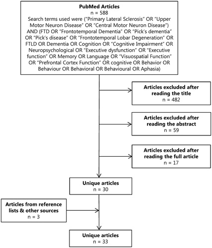
Figure 1. Summary of the results from our literature search. The search was performed using MEDLINE (via PubMed) on articles in English from July 1963 to January 2017 reporting on cognition and/or behaviour in PLS. Titles and abstracts were screened and relevant full text articles were retrieved. References were screened for additional studies and publications were also identified from the authors’ literature collections. When overlap between studies was expected, corresponding authors were contacted for further details. Since the inclusion of the search term “PLS” led to a high amount of irrelevant hits (e.g. partial least squares, post ligation syndrome, parathyroid lesions, etc.) it was not included in our list of search terms. We included studies on PLS in which dementia, cognition or behaviour was mentioned.

Figure 1. Summary of the results from our literature search. The search was performed using MEDLINE (via PubMed) on articles in English from July 1963 to January 2017 reporting on cognition and/or behaviour in PLS. Titles and abstracts were screened and relevant full text articles were retrieved. References were screened for additional studies and publications were also identified from the authors’ literature collections. When overlap between studies was expected, corresponding authors were contacted for further details. Since the inclusion of the search term “PLS” led to a high amount of irrelevant hits (e.g. partial least squares, post ligation syndrome, parathyroid lesions, etc.) it was not included in our list of search terms. We included studies on PLS in which dementia, cognition or behaviour was mentioned.

Figure 2. The initial axial T2 weighted MRI (A) showed left temporal lobe atrophy. The second scan made 2 years later (B) demonstrated bilateral temporal lobe and hippocampus atrophy (axial and coronal T2 weighted MRI), but more severe on the left side with a knife blade aspect of the temporal gyri while the general cerebral atrophy was limited (far right: sagittal T1 weighted MRI).

Figure 2. The initial axial T2 weighted MRI (A) showed left temporal lobe atrophy. The second scan made 2 years later (B) demonstrated bilateral temporal lobe and hippocampus atrophy (axial and coronal T2 weighted MRI), but more severe on the left side with a knife blade aspect of the temporal gyri while the general cerebral atrophy was limited (far right: sagittal T1 weighted MRI).

Table 1. Clinical characteristics of PLS-FTD patients.

Table 2. Studies on PLS patients that interrogated cognition and/or behaviour.

Table 3. Pooled weighted effect sizes and heterogeneity statistics of cognitive domains.

Table 4. Overview of neuroimaging findings in PLS.

Table 5. Overview of neuropathological findings in PLS.
Supplemental material

IAFD_Michael_et_al_Supplemental_content.zip

Download Zip (92.1 KB)