Figures & data
Figure 1 Proposed mechanisms underlying amyotrophic lateral sclerosis. (1) Nucleocytoplasmic transport defects including altered transport of RNA molecules and RNA-binding proteins. (2) Altered RNA metabolism. RNA-binding proteins including TDP-43 or FUS may become mislocalized in the cytoplasm leading to altered transcription and splicing. Stress granule dynamics are also affected. (3) Proteostasis is impaired with aggregating proteins including TDP-43 accumulating in the cytoplasm. There is evidence that the two main protein clearance pathways, autophagy and the UPS may be involved. (4) Impaired DNA repair: several ALS-linked genes including FUS, TARDBP, TAF15, SETX, and EWSR1 are involved in DNA repair. (5) Mitochondrial dysfunction resulting in the increased formation of reactive oxygen species (ROS) has been proposed as an initiating factor in ALS. Several ALS-linked proteins including SOD1, TDP-43, and FUS interact with mitochondria. (6) Axonal transport defects have been implicated in ALS pathogenesis. Neuropathological evidence has shown evidence of this including neurofilament accumulation and cytoskeletal disorganization. (7) Several ALS-linked genes including OPTN, VAPB, CHMP2B, and UNC13A are involved in vesicular transport. Impaired vesicular trafficking can lead to protein accumulation and golgi fragmentation which has been observed in ALS patients. (8) Neuroinflammation: the secretion of inflammatory proteins by activated microglia leads to the potentially neurotoxic activation of astrocytes, which may contribute to the death of neurons and oligodendrocytes. (9) Excitotoxicity: glutamate receptor overstimulation has been proposed to occur via several mechanisms including increased synaptic glutamate release, alterations to AMPA receptors and reduced clearance of glutamate by astrocytes. (10) Oligodendrocyte dysfunction may lead to reduced support for neurons. Changes in lactate production and transport via MCT1 have been implicated. Taken from Mejzini et al. (Citation37). Copyright © Mejzini, Flynn, Pitout, Fletcher, Wilton and Akkari. This is an open-access article distributed under the terms of the Creative Commons Attribution License (CC BY).
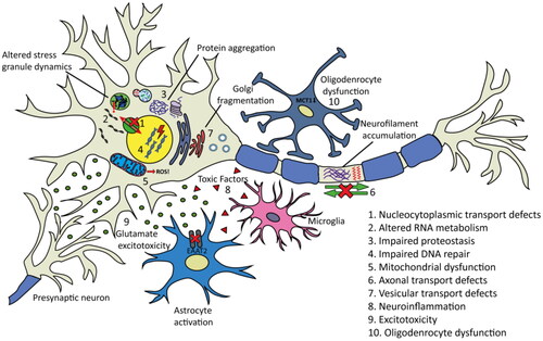
Figure 2 Timeline of amyotrophic lateral sclerosis diagnostic criteria and prognosis staging. Top timeline shows diagnostic criteria; bottom timeline shows prognostic staging. ALSFRS: ALS Functional Rating Scale; ALSFRS-R: ALS Functional Rating Scale-Revised; MiToS: Milano-Torino functional staging.
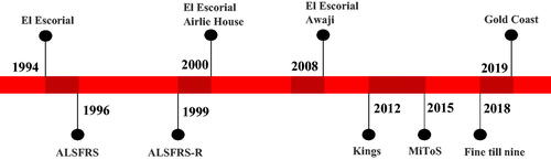
Table 1 Amyotrophic lateral sclerosis diagnostic criteria overview.
Table 2 Amyotrophic lateral sclerosis prognostic scoring and staging systems overview.
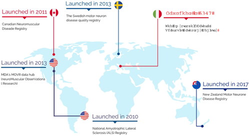
Figure 3 Map of worldwide registries in amyotrophic lateral sclerosis developed between 2010 and 2017. Adapted from Barbalho et al. (Citation2).