
Figures & data
Table 1. Optical imaging technology to improve tumour detection as adjuncts to white-light cystoscopy (adapted from Zlatev et al., 2015 [Citation15]).
Figure 1. Current and proposed future management pathway for patients with non-muscle-invasive bladder cancer. CT: computed tomography; MRI: magnetic resonance imaging; WLC: white-light cystoscopy; BLC: blue-light cystoscopy; OCT: optical coherence tomography; RS: Raman spectroscopy; CLE: confocal laser endomicroscopy; TURB: transurethral resection of the bladder; EORTC: European Organisation for Research and Treatment of Cancer; CUETO: Club Urológico Español de Tratamiento Oncológico; MMC: mitomycin C; BCG: bacille Calmette–Guérin; FU: follow-up; WLFC: white-light flexible cystoscopy; BLFC: blue-light flexible cystoscopy.
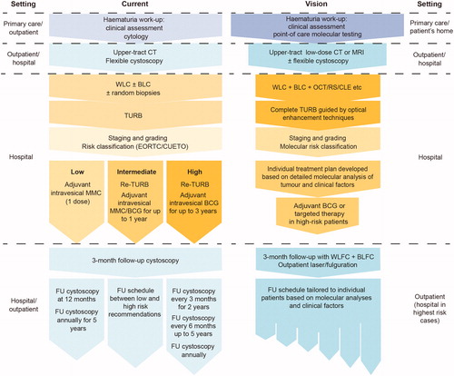
Table 2. Technological advances to improve diagnosis, treatment and follow-up of patients with non-muscle-invasive bladder cancer.