
Figures & data
Figure 1. Angiography of left inferior vesical artery, in Prostate Artery Embolization (PAE) treatment.
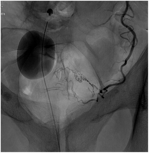
Figure 2. Cone-beam CT (CBCT) in coronal projection, showing contrast filling of prostate parenchyma and, thereby, correct catheter position prior to injection of particles in Prostate Artery Embolization (PAE) treatment.
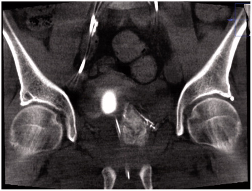
Figure 3. Angiography after Prostate Artery Embolization (PAE) treatment, showing occlusion of the inferior vesical artery.
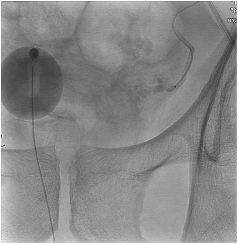
Table 1. Baseline data in patients (n = 37) treated with prostate artery embolization (PAE). Mean (SD) or N (%).
Table 2. Patients (n = 37) urinary catheter characteristics before and after treatment with prostate artery embolization (PAE).
Table 3. Changes in IPSS and QoL in patients without urinary catheter at baseline (n = 17) treated with prostate artery embolization (PAE).
Table 4. Characteristic in patients (n = 5) with clinical failure after treatment with prostate artery embolization (PAE).
Table 5. Complications in patients (n = 37) treated with prostate artery embolization (PAE).