
Figures & data
Table 1. Baseline patient, disease and tumor characteristics on MRI.
Table 2. TULSA for targeted ablation of PCa-lesions: Multimodality based volumetric correlations.
Figure 1. Location of the targeted lesions on baseline MRI. Note the red arrow pointing to the lesion on each image sequence. The first four patients had only one lesion, the last two patients also had a secondary lesion. Every lesion involved MRI-TBx-proven histologically significant PCa. On treatment planning, lesions were contoured with 5 mm of overlap when feasible, except near the NVB where a posterolateral 3 mm safety margin was ethically required for this treat-and-resect study, regardless of lesion coverage.
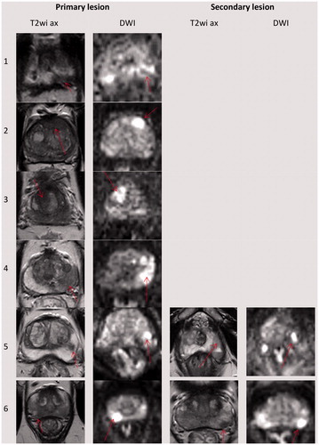
Figure 2. Lesion-targeted TULSA treatment (a) and 3-week follow-up (b) for patient number 2. (a) Intra-procedural MR images, with immediate post-treatment contrast-enhanced overlay images from active elements showing high spatial concordance between target volume (top row), 55 °C isotherm volume (cytocidal temperature, second row), thermal dose coverage (third row), and non-perfused-volume (NPV, fourth row). (b) Baseline imaging identified an anterior 3 cc PIRADS 5 lesion with a biopsy-concordant high-volume Gleason 3 + 3 PCa (MRI-TBx 4/4 positive cores, systematic biopsies 6 + 6 negative). Post-treatment, 1- and 3-week NPV and 3-week whole-mount hematoxylin-eosin-stained slide demonstrate high volumetric and morphometric concordance. Note that complete necrosis reached the capsule and thermal damage was well-confined. NPV covered the entire PIRADS 5 lesion and no vital cancerous tissue was reported in the vicinity of the targeted lesion. In final histopathology of the whole prostate specimen, only a posteroapical MRI-negative microfocus of vital histologically insignificant Gleason pattern 3 PCa was detected.
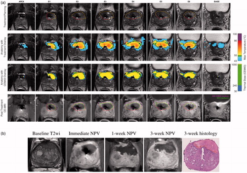
Table 3. TULSA for targeted ablation of PCa-lesions: The outcomes of treatment efficacy for targeted regions at 3 weeks