Figures & data
Figure 1. Representative images of the protein expression of versican in rat urinary bladder. (A) The tissue was stained without primary antibody against versican (negative control). (B and C) Application of the anti-versican antibody to the tissue resulted in immunoreactivity in the urothelium (U) and to a far lesser extent also in the detrusor muscle (D) and the lamina propria (Lp). Arrow points at microvilli-like structures at the urothelial surface. Scale bar: 100µm (A, B), 50µm (C).
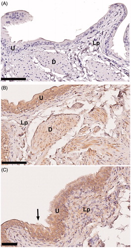
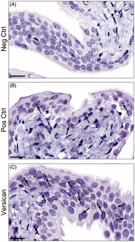
Figure 2. Representative images of the mRNA expression of versican in rat urinary bladder visualized using RNAscope® technology. (A) The urinary bladder was stained with DapB as a negative control. As expected no staining was seen. (B) Staining instead with polR2A as a positive control, generated a clear-cut positive reaction in the urothelial cells (arrows) and to a markedly lower extent in the remaining parts of the bladder. (C) The urinary bladder was stained for versican. Sparse, albeit clear, positive staining (arrows) was found in the urothelium, but also in scattered cells throughout the lamina propria, and the muscle cells (not shown in figure). Scale bar: 20 µm.