2,691
Views
14
CrossRef citations to date
0
Altmetric
Review

Quantitative analysis of nanoparticle transport through in vitro blood-brain barrier models

Article: e1143545 | Received 15 Dec 2015, Accepted 12 Jan 2016, Published online: 10 Mar 2016

Figures & data

Figure 1. Transwell system applied to measure the transport of nanoparticles across in vitro blood-brain barriers. A porous membrane, upon which the in vitro blood-brain barrier model is grown, separates two compartments. The nanoparticles are added to the upper compartment, and the number of nanoparticles that passes through to the lower compartment is measured.
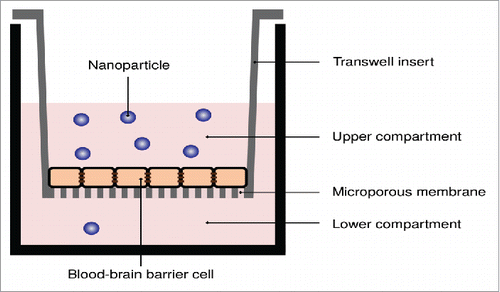
Figure 1. Transwell system applied to measure the transport of nanoparticles across in vitro blood-brain barriers. A porous membrane, upon which the in vitro blood-brain barrier model is grown, separates two compartments. The nanoparticles are added to the upper compartment, and the number of nanoparticles that passes through to the lower compartment is measured.

Figure 2. Potential issues with applying transwell systems to measure the transport of nanoparticles across in vitro blood-brain barriers.
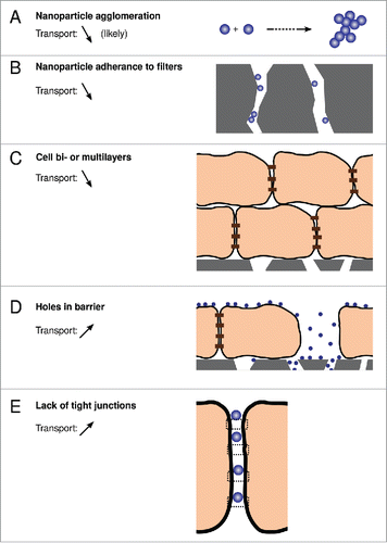
Figure 2. Potential issues with applying transwell systems to measure the transport of nanoparticles across in vitro blood-brain barriers.

Figure 3. Role of biomolecular corona in nanoparticle interactions with the blood-brain barrier. (A) Corona-covered nanoparticle interacting with cells of the barrier vs. (B) bare nanoparticle. Only the former situation is expected to occur in vivo.
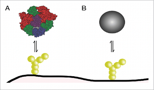
Figure 3. Role of biomolecular corona in nanoparticle interactions with the blood-brain barrier. (A) Corona-covered nanoparticle interacting with cells of the barrier vs. (B) bare nanoparticle. Only the former situation is expected to occur in vivo.

Figure 4. Indirect effects due to nanoparticle uptake in an in vitro blood-brain barrier. Despite the nanoparticles not being transported across the barrier (at least not to a significant degree), signaling takes place between the blood-brain barrier cells and astrocytic cells grown below them. Image adapted from ref. Citation86.
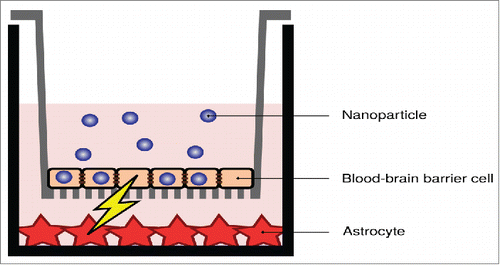
Figure 4. Indirect effects due to nanoparticle uptake in an in vitro blood-brain barrier. Despite the nanoparticles not being transported across the barrier (at least not to a significant degree), signaling takes place between the blood-brain barrier cells and astrocytic cells grown below them. Image adapted from ref. Citation86.