Figures & data
Figure 1. Mechanisms of OxPAPC-induced EC barrier protection. OxPAPC induces the activation of multiple signaling pathways that leads to the activation of Rap1 and Rac. The cytoskeletal remodeling facilitated by the cortical actin formation and assembly of tight junctions and adherens junctions proteins enhances lung endothelial barrier. In addition, EP4 receptor and lipoxin A4 also mediate the barrier protective effects of OxPAPC against LPS/TNFα-induced inflammation and lung injury in vitro and in vivo.
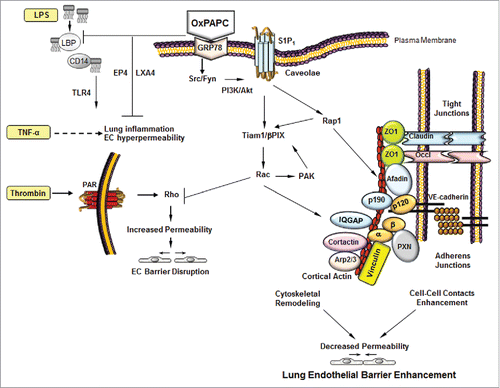
Figure 2. Generation of full length and fragmented oxidation products of 1-palmitoyl-2-arachidonoyl-sn-glycero-3-phosphorylcholine (PAPC).
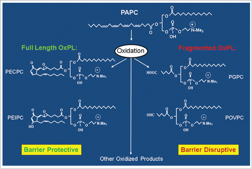
Figure 3. Metabolic pathway of sphingosine 1-phosphate (S1P) biosynthesis.
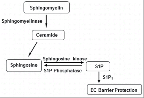
Figure 4. Biosynthesis of prostaglandins and thromboxanes from arachidonic acid.
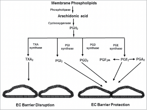
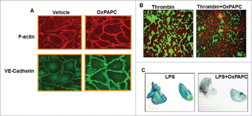
Figure 5. OxPAPC enhances endothelial barrier function and attenuates agonist-induced endothelial barrier dysfunction in vitro and in vivo. (A) HPAECs grown on coverslips were treated with OxPAPC (15 µg/mL, 30 minutes) and dual immunofluorescence staining with Texas-Red phalloidin and VE-cadherin was performed to monitor actin cytoskeleton and cell junction remodeling. The confocal images illustrated the barrier enhancing effects of OxPAPC as evidenced by increased F-actin and VE-Cadherin staining. (B) HPAECs grown on biotinylated gelatin substrate were pre-treated with OxPAPC (15 µg/mL, 30 minutes) followed by thrombin stimulation (0.5 Units/mL, 10 minutes) and cell permeability was visualized using FITC-avidin as a tracer. OxPAPC treatment protected thrombin-induced permeability as demonstrated by reduced green fluorescence that depicts areas permeable for FITC-avidin. (C) Intravenous injection of (1.5 mg/kg) of OxPAPC after 5 hours of LPS instillation (0.7 mg/kg, intratracheal) in mice protects against LPS-induced lung injury as detected by Evans blue staining of lung.