2,598
Views
0
CrossRef citations to date
0
Altmetric
Review

Maternal-fetal transmission of the zika virus: An intriguing interplay

ORCID Icon, &
Article: e1402143 | Received 08 Sep 2017, Accepted 30 Oct 2017, Published online: 25 Jan 2018

Figures & data

Figure 1. Schematic representation of cytotrophoblast differentiation and the invasion of the syncytiotrophoblast in the endometrium, contributing to the blastocyst nidation, from the sixth day after conception (p.c.). 6 days p.c. – Prelacunar stage where a mass of syncytiotrophoblastic cells invades the endometrium to become the decidua. Maternal-fetal exchanges are made by the syncytiotrophoblastic cells. 12 days p.c. – Lacunar stage / primary villi showing lacunas with maternal red cells in the syncytiotrophoblastic cell mass and sprouts of primary villi composed of cytotrophoblasts, facilitating maternal-fetal exchanges. 15 days p.c. – Stage of secondary villi where cytotrophoblastic secondary villi become more specialized and penetrate deeper into the mass of syncytiotrophoblastic cells and the lacunas become larger, facilitating even more maternal-fetal exchanges. 18 days p.c. – Stage of tertiary villi where the tertiary villi present primitive fetal mesenchymal and fetal vessels with fetal red blood cells. These specialized villi are bathed in the intervillous space, which is an expanse of the anterior lacunas, where the maternal blood facilitates maternal-fetal exchanges. The decidua is completely specialized, as is the placental bed. Detail: Cross-section of a fully specialized tertiary villus demonstrating the outermost syncytiotrophoblast continuous layer, the innermost cytotrophoblast layer, the fetal mesenchyme and the fetal vessels coated by the fetal endothelial cells. Hofbauer cells are found in the fetal mesenchyme.
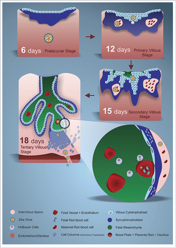
Figure 1. Schematic representation of cytotrophoblast differentiation and the invasion of the syncytiotrophoblast in the endometrium, contributing to the blastocyst nidation, from the sixth day after conception (p.c.). 6 days p.c. – Prelacunar stage where a mass of syncytiotrophoblastic cells invades the endometrium to become the decidua. Maternal-fetal exchanges are made by the syncytiotrophoblastic cells. 12 days p.c. – Lacunar stage / primary villi showing lacunas with maternal red cells in the syncytiotrophoblastic cell mass and sprouts of primary villi composed of cytotrophoblasts, facilitating maternal-fetal exchanges. 15 days p.c. – Stage of secondary villi where cytotrophoblastic secondary villi become more specialized and penetrate deeper into the mass of syncytiotrophoblastic cells and the lacunas become larger, facilitating even more maternal-fetal exchanges. 18 days p.c. – Stage of tertiary villi where the tertiary villi present primitive fetal mesenchymal and fetal vessels with fetal red blood cells. These specialized villi are bathed in the intervillous space, which is an expanse of the anterior lacunas, where the maternal blood facilitates maternal-fetal exchanges. The decidua is completely specialized, as is the placental bed. Detail: Cross-section of a fully specialized tertiary villus demonstrating the outermost syncytiotrophoblast continuous layer, the innermost cytotrophoblast layer, the fetal mesenchyme and the fetal vessels coated by the fetal endothelial cells. Hofbauer cells are found in the fetal mesenchyme.

Figure 2. Schematic representation of the possible human transmission routes of ZIKV: vector-borne and sexual. Following the possible routes of maternal-fetal transmission of ZIKV with their respective infectious consequences:- hematogenous, or transplacental routes, causing or not causing villitis, and reaching the fetus through the fetal vessels.- ascending leading to deciduites, chorionitis, amnionitis and funisitis and reaching the amniotic fluid, thus reaching the fetus.
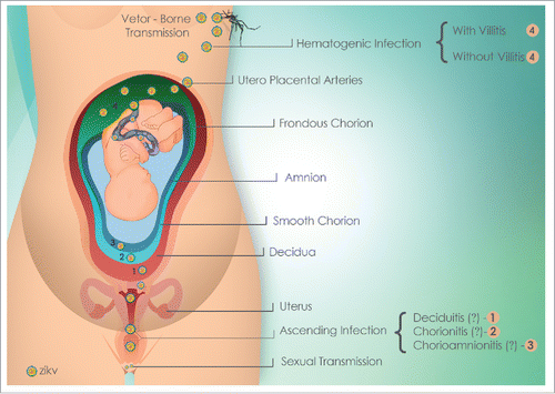
Figure 2. Schematic representation of the possible human transmission routes of ZIKV: vector-borne and sexual. Following the possible routes of maternal-fetal transmission of ZIKV with their respective infectious consequences:- hematogenous, or transplacental routes, causing or not causing villitis, and reaching the fetus through the fetal vessels.- ascending leading to deciduites, chorionitis, amnionitis and funisitis and reaching the amniotic fluid, thus reaching the fetus.

Reprints and Corporate Permissions

Please note: Selecting permissions does not provide access to the full text of the article, please see our help page How do I view content?

To request a reprint or corporate permissions for this article, please click on the relevant link below:

Academic Permissions

Please note: Selecting permissions does not provide access to the full text of the article, please see our help page How do I view content?

Obtain permissions instantly via Rightslink by clicking on the button below:

If you are unable to obtain permissions via Rightslink, please complete and submit this Permissions form. For more information, please visit our Permissions help page.