Figures & data
Figure 1. Characterization of PMN-derived EVs. (A-B) PMNs were stimulated with fMLF (1μM) to produce EVs. EVs were isolated by serial centrifugation and analyzed by transmission electron microscopy. (A) A representative EV with the size of exosomes (< 100 nm). (B) A representative microparticle/ectosome with the size of ∼600 nm. (C) PMNs (immunolabeled for CD11b, red and myeloperoxidase, green) release myeloperoxidase-containing EVs (shown by arrows) following adhesion to and migration across IECs (surface stain, blue).
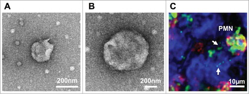
Table 1. A summary of miRNAs that have been shown to be transported by immune, epithelial, and endothelial cell-derived EVs and their contribution to cellular signaling and intestinal homeostasis.
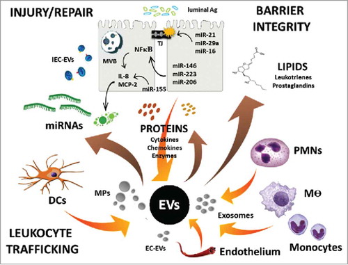
Figure 2. EV content and regulation of intestinal homeostasis. Schematic of EV release by various gut resident and recruited cells, as well as EV-mediated transport of proteins, lipids, and miRNAs to regulate cell function and intestinal homeostasis.