Figures & data
Figure 1. Expression of GPR40 and GPR120 in airway epithelial Calu-3 cells. Western blot analysis and immunofluorescence staining of GPR40 and GPR120 in Calu-3 cells are shown (n = 3–4). GPR40 and GPR120 in green (Alexa 488), nuclear content in blue (Hoechst), BF is brightfield.
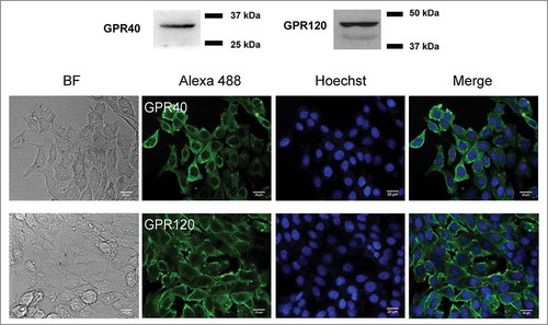
Figure 2. Effect of GPR40 on [Ca2+]i. Calu-3 cells were loaded with indo-1 and suspended in Ca2+-free buffers. After the indicated pretreatments (DC260126 (5 μM; GPR40 antagonist), AH7614 (100 μM; GPR120 antagonist), and U73122 (10 μM; PLC inhibitor) for an hour), GW9508 (5 μM) was added into bathing solutions during continual recording of indo-1 dual fluorescence (emitted at 405 nm and 490 nm). Representative tracings of indo-1 fluorescence ratio (F405/F490) and summary of the data are shown. Data are expressed as means of indo-1 fluorescence ratio (F405/F490) ±S.E.M. (n = 3–6). *** p < 0.001 compared with vehicle-treated group. ## p < 0.01; ### p < 0.001 compared with GW9508-treated group (one-way ANOVA).
![Figure 2. Effect of GPR40 on [Ca2+]i. Calu-3 cells were loaded with indo-1 and suspended in Ca2+-free buffers. After the indicated pretreatments (DC260126 (5 μM; GPR40 antagonist), AH7614 (100 μM; GPR120 antagonist), and U73122 (10 μM; PLC inhibitor) for an hour), GW9508 (5 μM) was added into bathing solutions during continual recording of indo-1 dual fluorescence (emitted at 405 nm and 490 nm). Representative tracings of indo-1 fluorescence ratio (F405/F490) and summary of the data are shown. Data are expressed as means of indo-1 fluorescence ratio (F405/F490) ±S.E.M. (n = 3–6). *** p < 0.001 compared with vehicle-treated group. ## p < 0.01; ### p < 0.001 compared with GW9508-treated group (one-way ANOVA).](/cms/asset/e2ed864c-cdd4-4dc5-9494-8eae11e6d468/ktib_a_1480741_f0002_b.gif)
Figure 3. Effect of GW9508 on tight junction assembly. Cells were cultured for 16 h in Ca2+-free media. TER of Calu-3 and 16HBE14o- cell monolayers was then measured after replacement of Ca2+-free media with media containing Ca2+ plus GW9508 at the indicated concentrations. (A) Effect of GW9508 on localization of ZO-1 in Calu-3 cells. After the indicated durations of incubation, cells were fixed and immunostained for ZO-1 (green) and nuclear content was stained with Hoechst (blue) (n = 4). (B) Effect of GW9508 on TER in Calu-3 cells. (Inset) Effect of GW9508 on TER in intact monolayers. (C) Expression of GPR40 and effect of GW9508 on TER in 16HBE14o- cells. After the indicated durations of incubation, TER was measured. Data are expressed as means of % of baseline TER ± S.E.M (n = 4–6). ** p < 0.01; *** p < 0.001 compared with vehicle-treated group (two-way ANOVA).
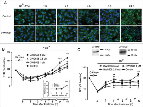
Figure 4. Roles of GPR40 and PLC in mediating GW9508-induced tight junction assembly. Cells were cultured for 16 h in Ca2+-free media. TER of Calu-3 cell monolayers was then measured after replacement of Ca2+-free media with the media containing Ca2+ plus vehicle or GW9508 (5 μM). (A) Role of GPR40. Cells were pretreated with GPR40 antagonists DC260126 (3 μM) or GW1100 (3 μM). (B) Role of PLC. Cells were pretreated with PLC inhibitor U73122 (20 μM). Data are expressed as means of % baseline TER ± S.E.M (n = 6). *** p < 0.001 compared with vehicle-treated group. # p < 0.05; ### p < 0.001 compared with GW9508-treated group. § p < 0.05; §§§ p < 0.001 compared between GW9508-treated and GW9508 + GW1100-treated groups (two-way ANOVA).
Figure 5. Involvement of AMPK in GPR40 stimulation-induced tight junction assembly. (A) Effect of metformin on tight junction assembly in Calu-3 cells. Cells were cultured for 16 hours in Ca2+-free media. TER of Calu-3 cells monolayers was then measured after replacement of Ca2+-free media with media containing Ca2+ plus vehicle, GW9508 (5 μM), or metformin (1.5 mM). Data are expressed as means of % baseline TER ± S.E.M (n = 4). ** p < 0.01; *** p < 0.001 compared between vehicle-treated and GW9508-treated groups. # p < 0.05 compared between vehicle-treated and metformin-treated groups (two-way ANOVA). (B) Involvement of AMPK in GW9508-induced tight junction assembly in Calu-3 cells. In the GW9508-treated group, cells were pretreated with vehicle or compound C (40 μM). Data are expressed as means of % baseline TER±S.E.M (n = 6). * p < 0.05; *** p < 0.001 compared with vehicle-treated group. ### p < 0.001 compared with GW9508-treated group (two-way ANOVA). (C) Summary of data at 8 h and 24 h. ** p < 0.05; *** p < 0.001 (one-way ANOVA). (D) Involvement of AMPK in GW9508-induced tight junction assembly in 16HBE14o- cells. * p < 0.05; ** p < 0.01 compared with vehicle-treated group. # p < 0.05; ## p < 0.01; ### p<0.001 compared with GW9508-treated group (two-way ANOVA).

Figure 6. GPR40 stimulation by GW9508 leads to AMPK activation. Western blot analyses of p-AMPK, AMPKα, and β-actin after treatment of Calu-3 cells with GW9508 at various times (A) and doses (B). Calu-3 cells were treated with GW9508 (5 μM) without or with DC260126 (3 μM) (C) and AH7614 (100 μM) (D) for 24 h before sample collection for western blot analysis. Data are expressed as mean of ratio of control (vehicle-treated group) ± S.E.M. (n = 4–5). * p < 0.05 compared with vehicle-treated group. ## p < 0.01 compared with GW9508-treated group (one-way ANOVA).
Figure 7. Involvement of CaMKKβ in AMPK activation following GPR40 stimulation. (A) Role of CaMKKβ in AMPK activation. Calu-3 cells were treated for 24 h with vehicle or GW9508 (5 μM) without or with pretreatment with CaMKKβ inhibitor STO-609 (5 μM) before sample collection for western blot analysis. Data are expressed as ratio of control (vehicle-treated group) ± S.E.M. (n = 4–5). * p < 0.05 compared with vehicle-treated group. # p < 0.05 compared with GW9508-treated group (one-way ANOVA). (B) Role of CaMKKβ in GPR40 stimulation-induced tight junction assembly. Cells were cultured for 16 h in Ca2+-free media. TER of Calu-3 cell monolayers was then measured after replacement of Ca2+-free media with media containing Ca2+ plus vehicle or GW9508 (5 μM) with or without pretreatment with STO-609 (5 μM). Data are expressed as means of % baseline TER ± S.E.M (n = 6). * p < 0.05; ** p < 0.01; *** p < 0.001 compared with vehicle-treated group. ## p<0.01 compared with GW9508-treated group (two-way ANOVA).