Figures & data
Figure 1. (A) Localization of sphingomyelin clusters was visualized with a sphingomyelin-binding toxin, lysenin. When cultured epithelial cells (EpH4 cells) were stained with green fluorescent protein (GFP)–lysenin, using anti-GFP antibody and the gold-conjugated secondary antibody, immunogold particles were present selectively at the microvilli in the apical membrane, suggesting that microvilli are specialized membrane structures where sphingomyelin clusters localize (Scale bar: 0.5µm). (B) PIP5Kbeta is recruited to the microvilli by its interaction with EBP-50, which in turn binds to podocalyxin-1 associated with sphingomyelin clusters.
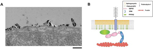
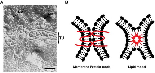
Figure 2. (A) A freeze fracture image showing tight junction strands in epithelial cells. MV: microvilli, TJ: tight junction. Scale bar, 200 nm. (B) Two models of tight junction. It is widely accepted that tight junctions are formed by the membrane protein, claudin (membrane protein model). The possibility that claudins or other tight junction membrane proteins help assembly and stabilization of a lipid-based strand structure is not completely denied (lipid model).