Figures & data
Figure 1. Embryonic tendons possess an organized actin cytoskeleton network, as well as cadherin-11 and N-cadherin cell-cell junctions. (a) The high cell density and an organized actin cytoskeleton network are visible in the midsubstance of E11 chick calcaneal tendons. Actin filaments (green) in embryonic tendon appear to form a continuous network between adjacent cells. (b) Cadherin-11 (red) and (c) N-cadherin (red) are present in E13 chick metatarsal tendons, but N-cadherin is localized to the exterior of the midsubstance of the tendon, while cadherin-11 appears in the interior of the midsubstance. Cell nuclei are labeled with blue. Figure 1A used with publisher’s permission from Schiele et al. 2015.Citation13 Scale bar = 10 μm. Figure 1B and 1C adapted with publisher’s permission from Richardson et al. 2007.Citation37
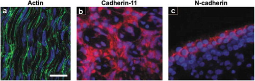
Figure 2. Longitudinal cryosections from equine CDET and SDFT tendons labeled for connexin-32 and connexin-43. (a) Connexin-32 (green) labeled in tendons from fetal, foal, and mature horses. (b) Connexin-43 (green) labeled in tendons from fetal, foal, and mature horses. Cell nuclei are labeled with red. Scale bar = 80 μm. Figure used with publisher’s permission from Stanley et al. 2007.Citation38.
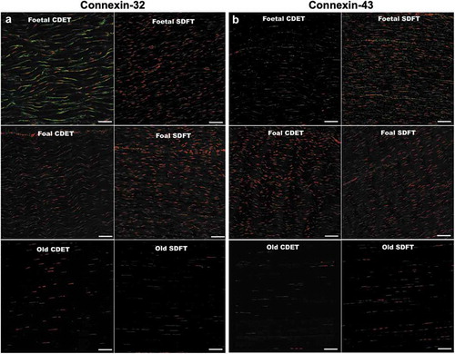
Table 1. Cell-cell junction proteins in developing tendon.
Table 2. Cell-cell junction proteins in adult tendon.
Figure 3. ZO-1 and claudin-1 are seen in a transverse section of E14 chick metatarsal tendon. Immunofluorescence staining showed the presence of an epithelium containing (a) ZO-1 (green) and (b) claudin−1 (red) tight junctions on the surface of the tendon. This epithelium may prevent tendon cell migration and tendinopathic adhesion formation. Figure used with publisher’s permission from Taylor et al. 2011.Citation76.
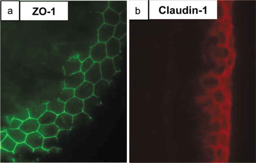
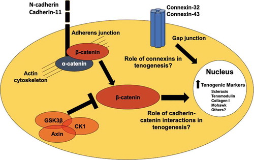
Figure 4. Potential cell-cell junction and cell signaling regulators of tenogenesis. N-cadherin and cadherin-11 are found in embryonic and adult tendon cells,Citation37,Citation81 and are regulated in tenogenically differentiating stem cells.Citation90 Cadherin-β-catenin coupling and β-catenin signaling may regulate tendon and stem cell behavior.Citation61,Citation62 Connexin-32 and connexin-43 are present in embryonic, postnatal, and adult tendon cells.Citation38,Citation66 Future studies are needed to understand what the roles these cell-cell junction proteins and β-catenin are playing in tenogenesis.