Figures & data
Figure 1. Effects of animal handling practices on baseline barrier function in uninjured small intestinal mucosa
(a) Acclimation status did not alter basal TEER in uninjured jejunal mucosa. N=8-11, P>.05 for acclimation effect by repeated measures mixed-effects analysis. (b) Environmental acclimation increased barrier permeability to 3H-mannitol in uninjured jejunal mucosa as compared to non-acclimated pigs in early ex vivo incubation. N=9, P=.045 for acclimation effect by two-way ANOVA, *P=.019 by Sidak’s multiple comparisons test.
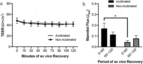
Figure 2. Combined effects of animal handing practices and acute ischemic injury on microscopic mucosal integrity in the small intestine
(a) Photomicrographs depicting representative histological views of epithelial integrity and microscopic tissue structure in acclimated and non-acclimated tissues subjected to control conditions, ischemic injury or injury and ex vivo recovery. Scale bar 100 µm. (b) Ischemia induced greater epithelial injury in non-acclimated pigs as compared to acclimated pigs. N=5-11, P=.013 for effect of acclimation by two-way ANOVA, ****P<.0001 by Sidak’s multiple comparisons test. (c) Data expressed as percent wound healing over the recovery period. N=5-11, P=.093 by Mann–Whitney test.
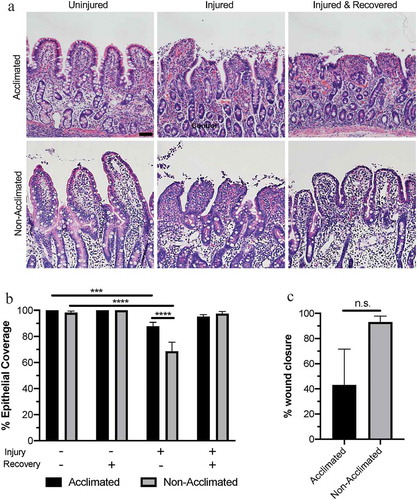
Figure 3. Combined effects of animal handing practices and acute ischemic injury on small intestinal barrier function
(a) Absolute TEER measurements increase over the ex vivo recovery period in ischemia-injured small intestine across groups, and higher resistance was detected in the non-acclimated group. N=7-11, P=.023 for the effect of acclimation by mixed effects model analysis. (b) Absolute mannitol flux showed increased macromolecular permeability in the early phase of ex vivo recovery in acclimated pigs. N=9-10, P= .317 for the effect of acclimation, P=.016 for interaction between acclimation and the recovery period by two-way ANOVA, **P<.01 by Sidak’s multiple comparisons test. (c) TEER measurements relative to control increase over the ex vivo recovery period in ischemia-injured small intestine across groups, and higher resistance was detected in the non-acclimated group. N=7-11, P=.010 for the effect of acclimation by mixed effects model analysis. *P<.05 and **P<.01 by Sidak’s multiple comparisons test. (d) Mannitol flux showed increased macromolecular permeability in the early phase of ex vivo recovery relative to control in non-acclimated pigs. This flux level decreased significantly in the later phase of ex vivo recovery. N=9-10, P= .0.038 for an interaction between acclimation and the recovery period by two-way ANOVA, *P<.05, **P<.01 by Sidak’s multiple comparisons test.
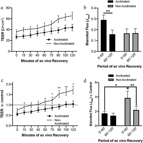
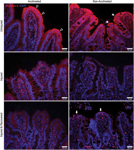
Figure 4. Combined effects of animal handing practices and acute ischemic injury on expression and localization of tight junction protein claudin-4 in the small intestinal mucosa
In uninjured mucosa, claudin-4 staining shows distinct localization at the cell borders, particularly at the apical membrane in non-acclimated pigs (top right panel, solid arrowheads) while the expression is more diffuse throughout the cytoplasm in acclimated pigs (top left panel, open arrowheads). With acute ischemic injury, expression appears to be broadly reduced with no particular localization to the cell membranes in both groups (middle panels). In mucosa recovered from ischemic injury, a small amount of returning claudin-4 expression is appreciated in the apical membranes of the newly restituted mucosal enterocytes of non-acclimated pigs localizing to the restoring tight junctions (bottom right panel, solid arrows). This pattern of staining is not visible in the acclimated pigs after ex vivo recovery (bottom left panel).