Figures & data
Figure 1. Schematic drawing of the BBB constituted by barrier type endothelial cell with TJ sharing the basement membrane with pericyte and the surrounding astrocyte endfeet. Created with BioRender.com
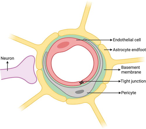
Figure 2. Schematic drawing of the BCSFB constituted by epithelial cells of choroid plexus with TJs, which secrete CSF derived from plasma in blood capillary without barrier properties into the ventricular space lined with ependymal cells. Created with BioRender.com
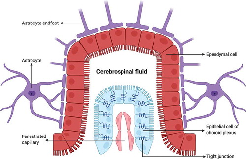
Figure 3. An electron micrograph from our image archive showing a pericyte (p) partly investing the endothelial cells of blood capillaries. Note that both endothelial cells and the pericyte are embedded in the basement membrane marked by extravasated electron-dense horseradish peroxidase tracer accumulation (arrows) owing to BBB disruption. A: astrocyte endfoot
Figure 4. A light micrograph from our image archive showing astrocytes labeled by immunostaining for GFAP in the hippocampal region of the brain. Note the microvessels almost entirely surrounded with astrocyte endfeet (arrows)
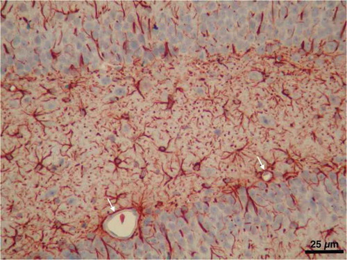
Figure 5. Schematic drawing of the junctional complex between barrier type endothelial cells of the brain, which controls the trafficking of substances through the paracellular pathway. The major proteins comprising TJs and adherens junctions and their linkage to the actin cytoskeleton are illustrated. Created with BioRender.com
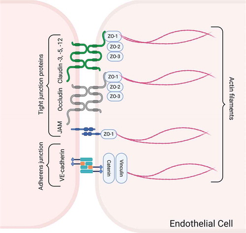
Figure 6. The schematic drawing of various routes of transcellular transport across barrier type endothelial cells in the brain. A: passive diffusion, B: efflux transport, C: carrier-mediated transport, D: receptor-mediated transport, E: adsorptive-mediated transport, F; cellular transport. Created with BioRender.com
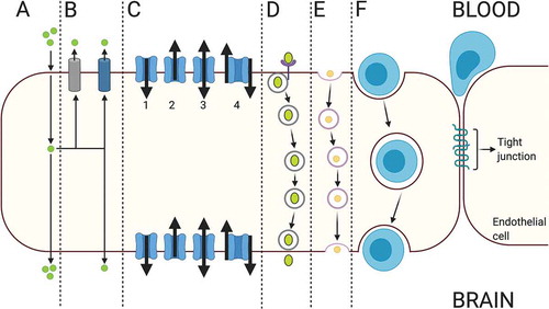
Figure 7. An electron micrograph from a previous study from our research group (reproduced from Ref. # 96 with permission from Elsevier Science) showing a capillary from the hippocampus region of the brain of a rat with cortical dysplasia exposed to febrile seizures. Note a conglomerate of caveolar vesicles within a cargo (arrow in the inset) in the cytoplasm of a brain capillary endothelial cell and the intensive pericapillary edema with swollen astrocyte endfeet