Figures & data
Figure 1. Epithelial barrier and its connections. Tight Junctions form an apical junction complex by forming a ring in the apicolateral region of the intestinal, skin and airway barrier. AJs in the lower region regulate the apical-basolateral membrane. TJs and AJs bind to the actin cytoskeleton via ZO-1, ZO-2, ZO-3. They are desmosomes that connect epithelial cells and disperse on the lateral surfaces. a-Catenins do not bind directly to cadherins; It binds to actin via ZO-1. Cadherins can become functional by interacting with catenin proteins.Citation9
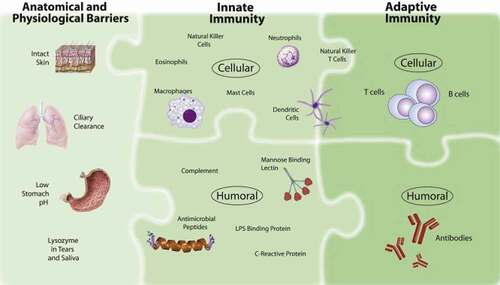
Figure 2. Immune system: (1) Anatomical and physiological barriers; (2) innate immune system elements; (3) Adaptive immune system elements. The harmony between these systems ensures the correct functioning of the immune mechanism and the effective functioning of the immune system.Citation8
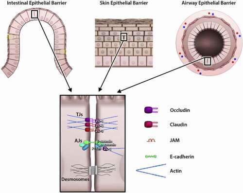
Figure 3. Microstructures of allergens and effector regions in diagnostic tests. IgE reactions are shown with different diagnostic principles and allergological tests. It indicates the fraction of total IgE antibody sIgE in serum specific for certain allergens. sIgEs show a particular sensitivity to the allergen to which they are specific. It responds in different ways in different effector organs with different diagnostic methods.Citation14
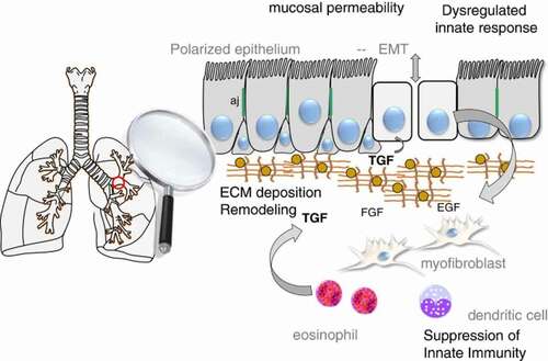
Figure 4. Schematic view of epithelial mesenchymal transition in the airway. It has an important place in the epithelium in asthma. In the relationship expressed as the epithelial mesenchymal unit, signals are sent through the mesenchymal in the epithelial sub-epithelial environment. It ensures the integrity of this unit of growth. Epithelial growth factor (EGF), Fibroblast growth factor (FGF), cytokines (such as IL-6, IL-1p, TGF-beta) release increases.Citation32
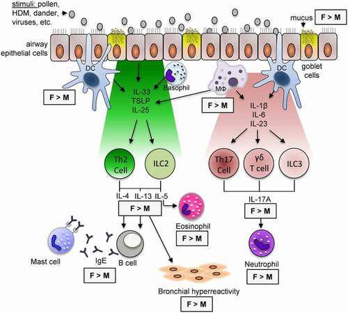
Figure 5. Triggers and mechanisms in airway changes, with gender differences. In asthma-related airway inflammatory diseases, the affected areas and the responses shown may vary according to gender. There are also gender differences in type-2 and IL-17A mediated airway inflammation.Citation23