Figures & data
Figure 1. Schematic of cross-section through the glomerular filtration barrier (GFB) showing key components of the barrier, including the fenestrated endothelia, the podocyte foot processes separated by slit diaphragms and the GBM. Not shown is the glycocalyx covering the endothelial cells and podocyte foot processes.Citation1 The small images on the right give a ‘top down’ view of the interdigitating foot processes of adjacent podocytes (top) and the appearance of fenestra in the endothelial cells (bottom).
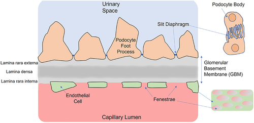
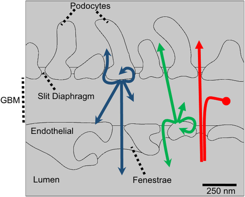
Figure 2. Solutes within the GBM may be from a number of sources and have one of several destinations. The role of GFB as a filter focuses on solutes sourced from the blood plasma (shown by red arrows). These molecules may pass through the GBM, accumulate in the GBM (potentially leading to clogging) or are excluded (e.g. by anion exclusion). Molecules synthesized by either the podocytes (blue arrows) or endothelial cells (green arrows) may potentially leave the GBM via the fenestra or the slit diaphragms. Alternatively, they may spend time in the GBM, contributing to the GBM ECM until they are degraded or transported away. In addition, cross-talk (paracrine signaling) between podocytes-and endothelial cells and autocrine signaling is also possible, though not the focus of the current review.