Figures & data
Table 1. Bacteria and medium used in this study.
Figure 1. Lactobacillus delbrueckii subsp. bulgaricus 2038 and Streptococcus thermophilus 1131 improved the intestinal barrier dysfunction by tumor necrosis factor (TNF)-α and interferon (IFN)-γ.
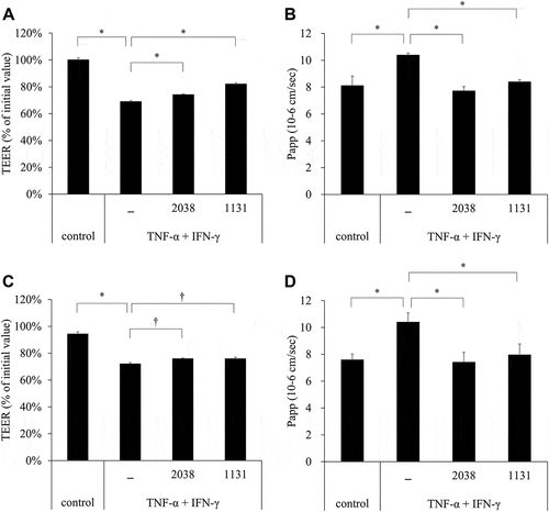
Figure 2. L. bulgaricus 2038 and S. thermophilus 1131 suppressed the decrease in the gene expression levels of tight junctions (TJs) by TNF-α and IFN-γ.
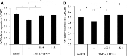
Figure 3. L. bulgaricus 2038 and S. thermophilus 1131 suppressed the destruction of TJ proteins by TNF-α and IFN-γ.
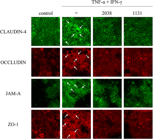
Figure 4. L. bulgaricus 2038 and S. thermophilus 1131 promoted the assembly of TJ proteins in a calcium switch assay.
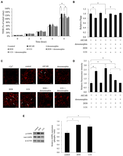
Figure 5. L. bulgaricus 2038 and S. thermophilus 1131 increased the expression of p-AMPK.
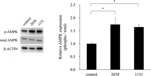
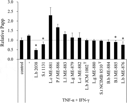
Figure 6. Protective effect on FD-4 permeability increased by TNF-α and IFN-γ was strain-dependent.