Figures & data
Figure 1. Scanning electron microscopy images of chitosan beads (a, c, e), immobilized lipase in chitosan beads (b, d, f) with different magnifications (130×, 1000×, 10,000×, respectively).
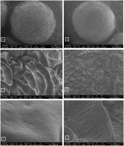
Figure 2. Optimization of chitosan concentration used immobilization procedure.
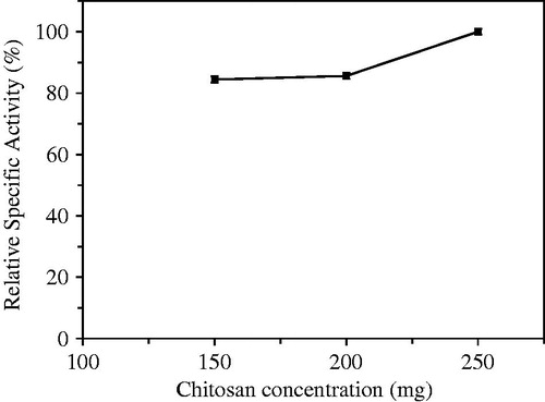
Figure 3. Optimization of tripolyphosphate (TPP) concentration used as multivalent covalent counter ion in immobilization procedure.
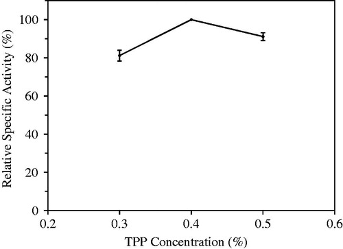
Figure 4. Optimization of buffer pH prepared tripolyphosphate (TPP) used as multivalent covalent counter ion.

Figure 5. Optimization of laurel lipase amount used in immobilization procedure.
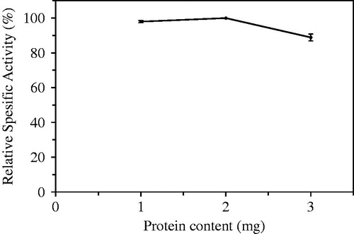
Figure 6. Optimization of bead size prepared in lipase immobilization.
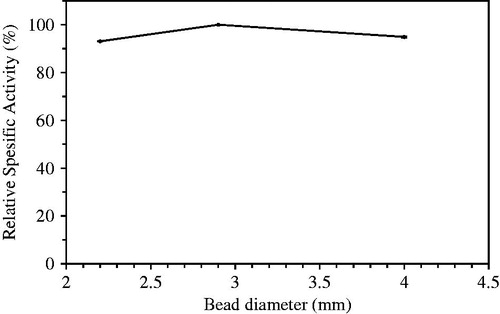
Figure 7. Optimization of bead amount used in assays of hydrolytic activity catalyzed laurel lipase.
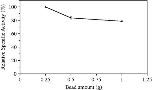
Figure 8. Optimum pH of immobilized laurel lipase.
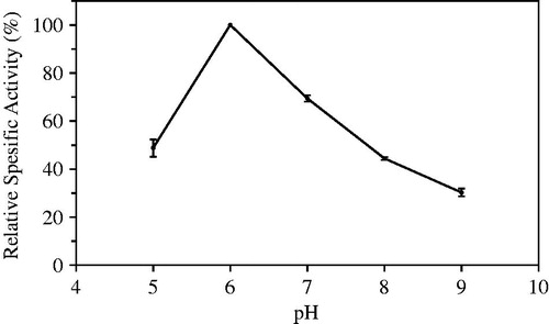
Figure 9. Optimum temperature of immobilized laurel lipase.
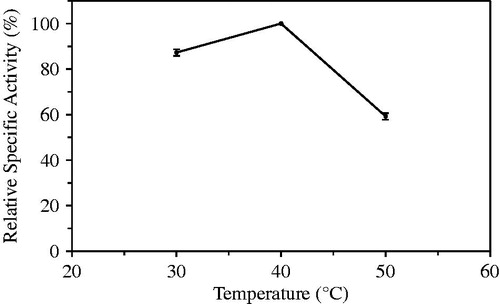
Figure 10. Thermal stability of immobilized laurel lipase.
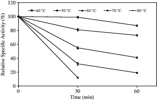
Figure 11. Lineweaver-Burk graph of immobilized and free laurel lipase.
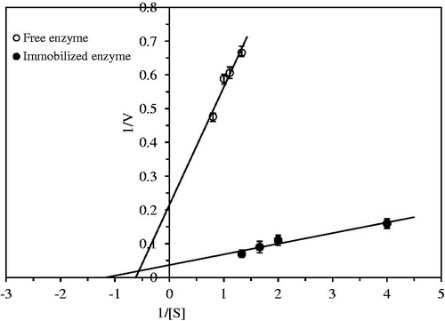
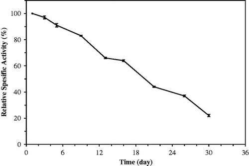
Figure 12. Storage stability of immobilized laurel lipase.