Figures & data
Figure 1. Photograph showing color changing (A) aqueous leaf extract of Catharanthus roseus (B) changing color from yellowish to reddish brown after adding 2 mM AgNO3 and exposing to heat at 70 °C for 3 min. (C) 2 mM AgNO3 only.
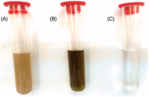
Figure 2. UV-vis absorption spectra of reduction of silver ions to silver nanoparticles.
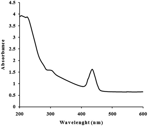
Figure 3. XRD pattern of silver nanoparticles formed after reaction with Catharanthus roseus leaf extract.
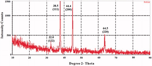
Figure 4. The FTIR spectra of silver nanoparticles.
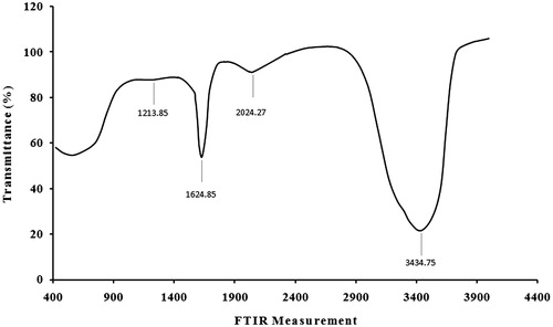
Figure 5. FM image of silver nanoparticles film showing uniformly distributed nanoparticles and some agglomeration.
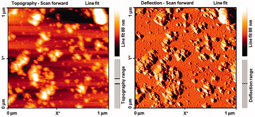
Figure 6. DPPH-free radical scavenging activity of silver nanoparticles formed after reaction with Catharanthus roseus leaf extract. Results are expressed as percentage decrement of absorbance at 517 nm with respect to control. Each value represents the mean ± SD of three experiments.
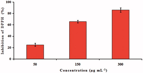
Figure 7. Antimicrobial activity assay of silver nanoparticles against different pathogens by the well diffusion method. (A) Amoxicillin/or fluconazole (B) Catharanthus roseus leaf extract, (C) silver nitrate, (D) synthesized silver nanoparticles at 100 μg mL−1, and (E) synthesized silver nanoparticles at 200 μg mL−1. Antibiotic amoxicillin at concentration 30 μg mL−1 was used as a control for all tested bacteria while, fluconazole at concentration 5 μg mL−1 was used as a control for Candida albicans.
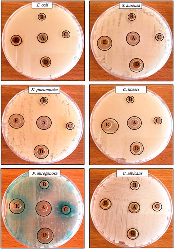
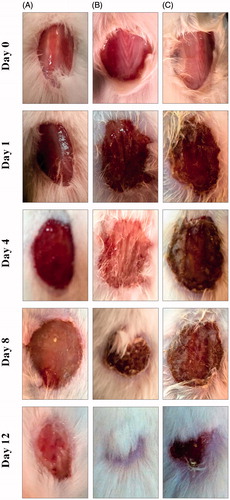
Figure 8. Photographs of wounds from animals elucidation on different days of (A) negative control, (B) silver nanoparticles, and (C) Catharanthus roseus leaf extract-treated mice.