Figures & data
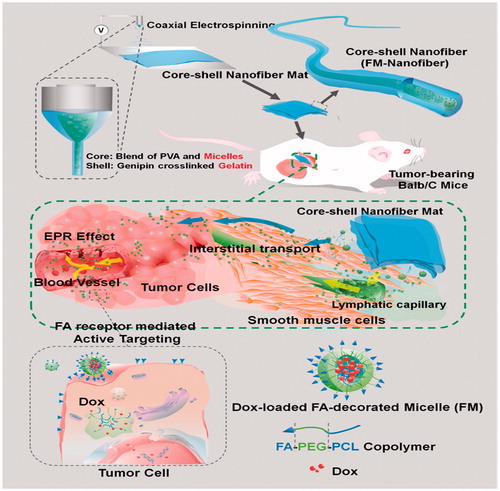
Figure 1. Drug incorporation techniques (Goonoo et al. Citation2014).
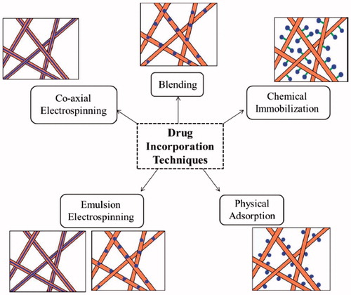
Figure 2. Schematic illustrations of the fabrication of the implantable active-targeting micelle-in-nanofiber device (FM-Nanofiber) and the delivery process of these Dox-loaded micelles (FM) from nanofiber matrix to tumor tissues and finally to tumor cells (Yang et al. Citation2015).