Figures & data
Figure 1. (a) Growth curve of F. oxysporum (PTCC 5291) fungus in MGYP medium at 28 °C; (b) UV–visible spectra of colloidal AgNPs originated from culture medium of F. oxysporum fungus at mid logarithmic (I), late logarithmic (II), and stationary (III) phases; (c) Nitrate reductase enzyme activity in biomass-free filtrates of F. oxysporum acquired at different growth phases.
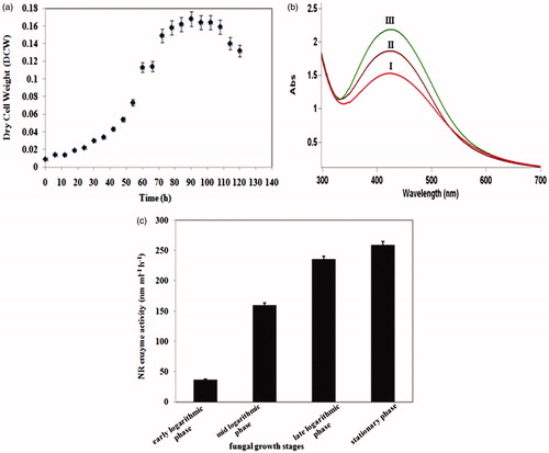
Figure 2. UV–visible spectra of colloidal AgNPs synthesized using cell-free filtrates of F. oxysporum fungus which was grown at (a) 23 °C, (b) 28 °C, and (c) 33 °C after 24 h (I) and 48 h (II).
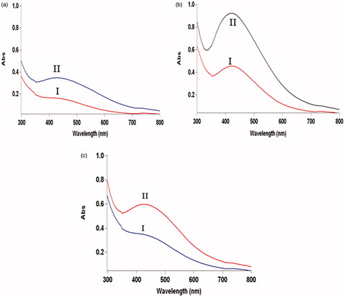
Figure 3. UV–visible spectra of colloidal AgNPs produced using cell-free filtrates of F. oxysporum which incubated in (a) MGYP and (b) PDB media in the presence of light; after 24 h(I), 48 h (II), and 72 h (III); (c) MGYP and (d) PDB media in absence of light; after 48 h (II) and 72 h (III); (e) DLS analyses of colloidal AgNPs generated by mixing of AgNO3 solution and cell-free filtrates of F. oxysporum cells which cultured in presence of light in (I) MGYP and (II) PDB media.
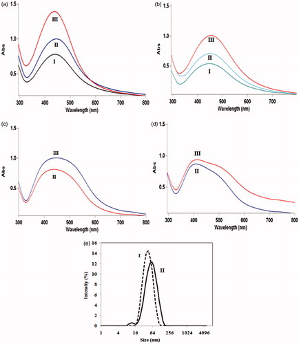
Table 1. Comparison of total proteins and NR enzyme secretions in cell-free filtrates of F. oxysporum cultured in MGYP and PDB media both in the presence and in the absence of light.
Figure 4. UV–visible spectra of colloidal AgNPs in reaction solutions containing 1:1 volume ratio of AgNO3 solutions and cell-free filtrates originated from F. oxysporum cells incubated at (a) MGYP medium, (b) modified medium after 12 h (I), 24 h (II), 36 h (III), 60 h (IV), and 72 h (V); (c) DLS analysis of colloidal AgNPs generated by the mixtures of AgNO3 and cell-free filtrates of F. oxysporum incubated in (a) MGYP medium; (b) modified medium containing potassium nitrate; (d) SEM image of synthesized AgNPs in reaction mixture containing silver nitrate, and cell-free filtrate of incubating F. oxysporum in modified medium containing nitrate source.
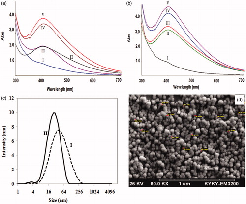
Figure 5. UV–vis analysis of colloidal AgNPs synthesized using mixtures of silver nitrate solutions and filtrates of submerged F. oxysporum biomass in ammonium sulphate (I); ammonium nitrate (II); distilled water (III); 25 mM potassium nitrate (IV); 50 mM potassium nitrate (V); after 72 h (reaction completion).
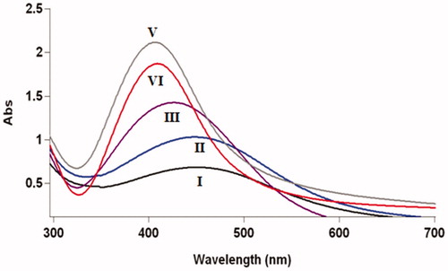
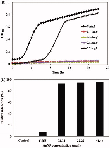
Figure 6. (a) Growth curves of E. coli in LB culture media supplemented with 5.55, 11.11, 22.22, and 44.44 mg/l AgNPs synthesized using cell-free filtrate of F. oxysporum; (b) bacterial growth inhibition after 18 h treatment with different concentrations of AgNPs.