Figures & data
Table. 1 Primers used in real-time RT-PCR.
Figure 1. SEM images. (A and B) Dex-embedded PEO nanofibers composited to PCL (PEO/Dex-PCL) at two magnifications. (C) MSCs seeded PEO/PCL and (D) PEO/Dex-PCL at day 7 and (E) MSCs seeded PEO/PCL and (F) PEO/Dex-PCL at day 21.
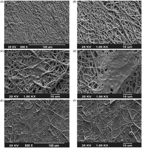
Figure 2. Contact angle measurements to determine hydrophilic properties of PCL, PEO/PCL, and PEO/Dex-PCL nanofibrous scaffolds.
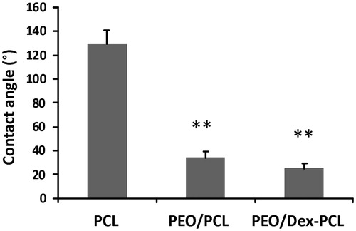
Figure 3. The profile of released dexamethasone from PEO/Dex-PCL nanofibrous scaffold during one week.
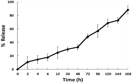
Figure 4. Viability of adipose tissue-derived MSCs seeded on TCPS-Dex, PEO/PCL, PEO/Dex-PCL, and TCPS as control during 5 days culture period (Significant difference between the groups at *P < 0.05).

Figure 5. (A, B) Morphology of MSCs at passage 3 before use for differentiation at two magnifications (10× and 40×). (C–H) MSC surface markers evaluation. (I, J) Morphology of differentiated MSCs 21 days after culture under medium (10× and 40×).
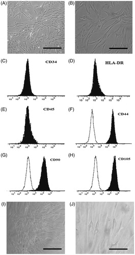
Figure 6. Glycosaminoglycan (GAG) staining of the extracellular matrix of differentiated cells cultured on TCPS at (B) days 14 and (D) 21, PEO/PCL at days (F) 14 and (H) 21 and PEO/Dex-PCL at days (J) 14 and ( L) 21, (A, C, E, G, I, and K) DAPI-stained nanofibers at days 14 for each groups, magnifications 10×.
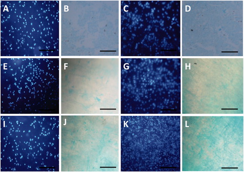
Figure 7. Relative expression of chondrogenic genes on 7, 14, and 21 days in MSCs cultured on PEO/PCL-DM, PEO/Dex-PCL-BM, PEO/Dex-PCL-DM, and TCPS-DM as control, (Significant difference between the groups at *P < 0.05).
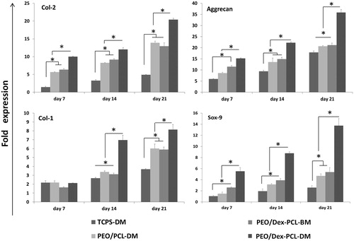
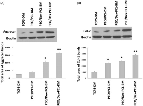
Figure 8. Western blot results: (A) Aggrecan, and (B) Collagen type 2 on day 21 in MSCs cultured on PEO/PCL-DM, PEO/Dex-PCL-BM, PEO/Dex-PCL-DM, and TCPS-DM as control, (Significant difference between the groups at *P < 0.05, **P < 0.01).