
Figures & data
Figure 1. Effect of 90 min sustained hemorrhagic shock on intracellular pCO2 and effects of different transfusion fluids. From Bian and Chang [Citation13] with copyright permission.
![Figure 1. Effect of 90 min sustained hemorrhagic shock on intracellular pCO2 and effects of different transfusion fluids. From Bian and Chang [Citation13] with copyright permission.](/cms/asset/27161004-9d75-42a3-a283-2148995c835e/ianb_a_1293676_f0001_c.jpg)
Figure 2. Effects of different transfusion fluids on the recovery of ST elevation in 90 min sustained hemorrhagic shock rat model. From Bian and Chang [Citation13] with copyright permission.
![Figure 2. Effects of different transfusion fluids on the recovery of ST elevation in 90 min sustained hemorrhagic shock rat model. From Bian and Chang [Citation13] with copyright permission.](/cms/asset/0dcfae26-5aea-4a96-ba0f-99d27a7adbe5/ianb_a_1293676_f0002_c.jpg)
Figure 3. Hematoxylin and eosin staining of intestine tissue sections (200×) of Shed blood reperfusion group after 90 min ischemia and 1 h resuscitation.
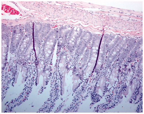
Figure. 4 Hematoxylin and eosin staining of intestine tissue sections (200×) of PolyHb reperfusion after 90 min ischemia and 1 h resuscitation.
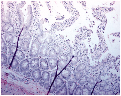
Figure 5. Hematoxylin and eosin staining of intestine tissue sections (200×) of Poly-[SFHb-SOD-CAT-CA] reperfusion after 90 min ischemia and 1 h resuscitation.
![Figure 5. Hematoxylin and eosin staining of intestine tissue sections (200×) of Poly-[SFHb-SOD-CAT-CA] reperfusion after 90 min ischemia and 1 h resuscitation.](/cms/asset/1753ba0d-0d10-4f71-a5f3-987fcd45300f/ianb_a_1293676_f0005_c.jpg)
Figure 6. Different type of Poly-[Hb-SOD-CAT-CA].
![Figure 6. Different type of Poly-[Hb-SOD-CAT-CA].](/cms/asset/c5b07303-22e8-426a-b08d-c6f0ed88e62a/ianb_a_1293676_f0006_c.jpg)
Table 1. Comparison of PolyHb, Poly-[Hb-SOD-CAT] and Poly-[Hb-SOD-CAT-CA] with RBC.