Figures & data
Figure 1. Spinal cord injury in rats. (A) Spinal cord contusion injury resulted in a loss of tissue structure within the central region of the spinal cord by 2 week post-injury, the pathological tissue was formed after injury and dropped out when tissue sliced. (B) Evaluation of motor function score. Trauma caused a significant change in motor function score as compared with the control group values (normal).
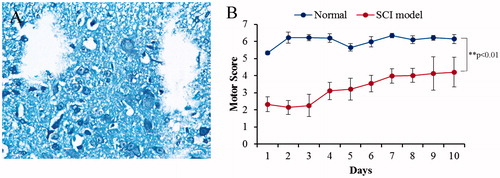
Figure 2. Biological characterization of exosome from CSF between SCI and normal. Exosomes exhibit a cup- or round-shaped morphology under ETM, with a size ranging from 50 to 80 nm. Meanwhile, these exosomes expressed exosomal surface marker proteins, including CD9, CD63, CD81, Alix and Tsg10.
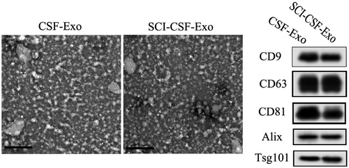
Figure 3. Neurons makers were detected in rat neurons derived from spine cord. Immunoflourescence staining and flow cytometry assay indicated that rat neurons expressed specific genes, including β-III-tubulin and MBP.
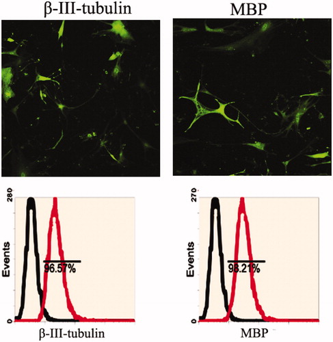
Figure 4. Exosome promotes proliferation of rat neurons. (A) Cell viability in 1- to 8-day-cultured rat neurons after exosome derived CSF treatment via MTT assay. (B) Cell proliferation in 1- to 8-day-cultured rat neurons after exosome derived CSF treatment via BrdU labeling indicating increased proliferation with SCI-CSF-exosome treatment. (C) Western blot of activation of ERK1/2. Increased expression level of ERK1/2 after SCI-CSF-exosome treatment, but no significant changes after normal CSF-exosome treatment compared with the control.
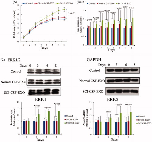
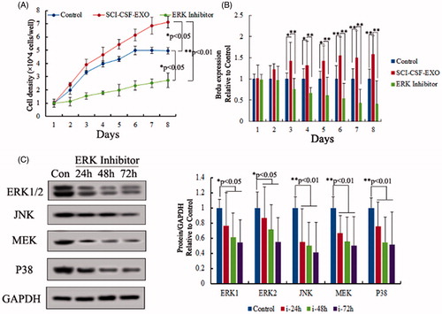
Figure 5. Role of ERK pathway in neurons proliferation. (A) Cell viability in 1- to 8-day-cultured rat neurons after SCI-CSF-exosome, ERK pathway inhibitor treatment respectively via MTT assay. (B) Cell proliferation in 1- to 8-day-cultured rat neurons after SCI-CSF-exosome treatment, ERK pathway inhibitor treatment via BrdU labeling. The results indicated ERK pathway inhibitor was a blocker for rat neurons after SCI-CSF-exosome treatment. (C) Expression level of ERK pathway members after inhibitor treatment, the results indicating expression level of ERK pathway members, including ERK 1/2, JNK, MEK and P38, showed a time-lapse increase.