Figures & data
Table 1. Phytochemical screening of C. fistula fruit pulp extract.
Figure 1. The variation of colour changes in tested compounds (A) before and (B) after the process of reduction of Ag + to Ag0 nanoparticles. (C) UV–Vis absorption spectra of AgNPs synthesized using aqueous pulp extract of C. fistula after 24 h.
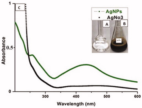
Figure 2. The FTIR spectra of AgNPs biosynthesized by C. fistula aqueous pulp extract.
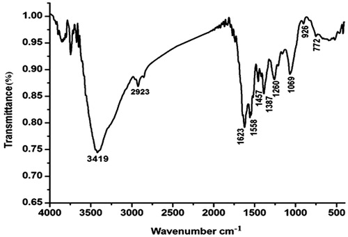
Figure 3. (A) SEM, (B) TEM images of AgNPs synthesized by reducing 3 mM AgNO3 using aqueous pulp extract of C. fistula, (C) EDX results of the AgNPs formation.

Figure 4. XRD patterns of synthesized AgNPs from aqueous pulp extract of C. fistula.
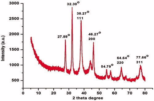
Table 2. Larval and pupal toxicity using fruit pulp aqueous extract of C. fistula against Ae. albopictus.
Table 3. Larval and pupal toxicity using fruit pulp aqueous extract of C. fistula against Cx. pipiens pallens.
Table 4. Larval and pupal toxicity of silver nanoparticles synthesized using C. fistula fruit pulp extract against Ae. albopictus.
Table 5. Larval and pupal toxicity of silver nanoparticles synthesized using C. fistula extract against Cx. pipiens pallens.
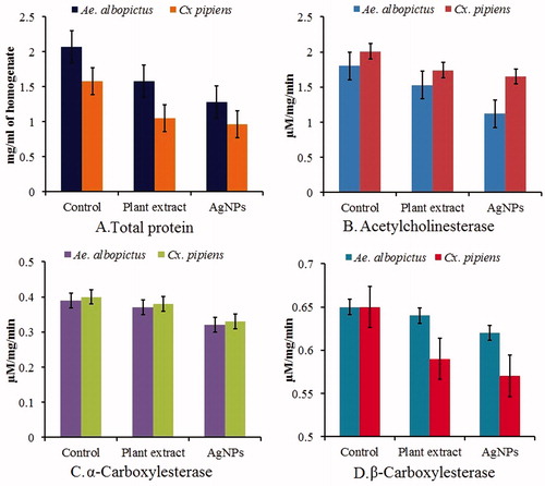
Figure 5. Biochemical analysis of aqueous plant extract and AgNPs treated (Test) and untreated (Control) of fourth instar larvae of vector mosquito Ae. albopictus and Cx. pipiens pallens showing differential response on total protein, acetylcholine esterase, α- and β- carboxylesterase.