Figures & data
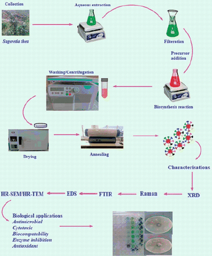
Figure 1. Study design.
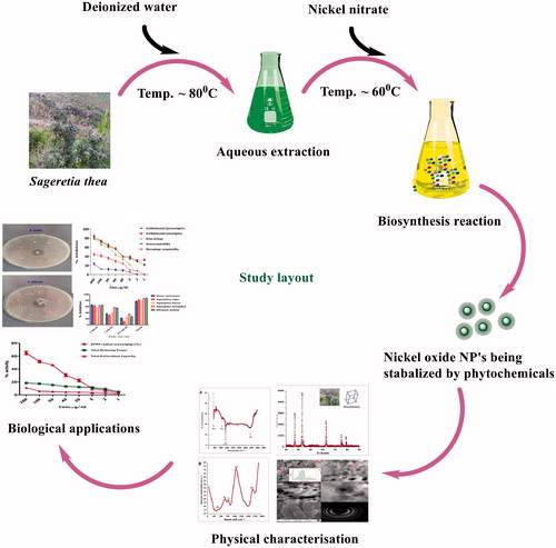
Figure 2. XRD analysis. (A): Typical XRD pattern of NiO annealed at 500 °C; (B): size calculation according to Scherer approximation.
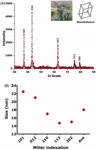
Figure 3. Morphological investigations using HR-TEM/HR-SEM; (A/B/C/D): size distribution of NiO nanoparticles; (E): HR-TEM image; (F): HR-SEM; (G): SAED pattern.
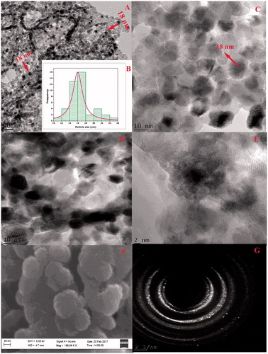
Figure 4. (A) Typical ATR-FTIR of NiO nanoparticles; (B) their Raman spectra.
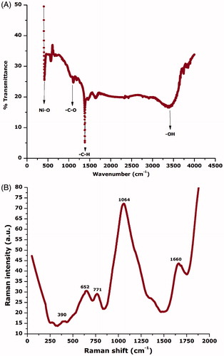
Figure 5. Elemental composition using energy-dispersive spectroscopy (EDS).
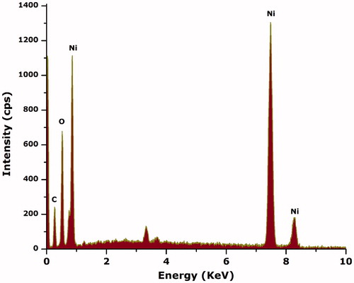
Figure 6. Stability of NiO nanoparticles.
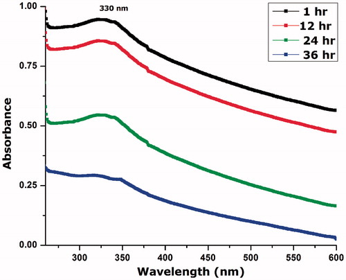
Figure 7. (A) Antibacterial activities of NiO nanoparticles without UV illumination; (B) with UV illumination.
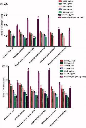
Figure 8. Antifungal potential of bioinspired NiO nanoparticles.
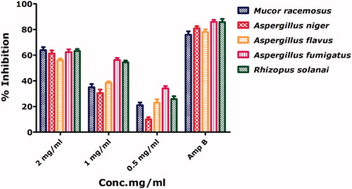
Table 1. MIC calculations against gram positive and gram negative bacterial strains.
Figure 9. Assessment of the cytotoxicity of the bioinspired NiO nanoparticles.
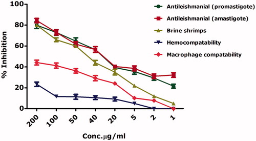
Figure 10. Schematic representation of the cytotoxic properties of bioinspired NiO as reported in literature; (A): ROS generation; (B): Ni++ release from NiO; (C): membrane damage by interference of membrane proteins with ROS or with their interference with surface defected NiO; (D): interference of NiO nanoparticles/ROS/Ni++ with nuclear material; (E): their interference with proteins; (F): entrance to mitochondrial to generate further ROS; (G): adherence to the membranes and pores.
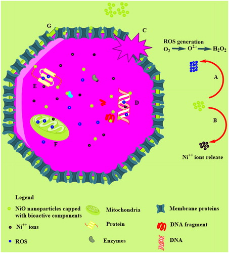
Table 2. IC50 calculations using table curve software.
Figure 11. Antioxidant activities of bioinspired NiO.
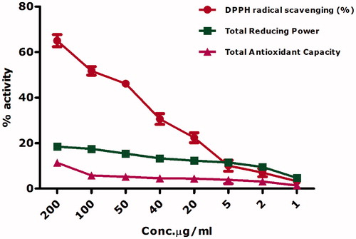
Figure 12. Ascorbic acid calibration curve.
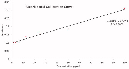
Figure 13. (A): Protein kinase inhibition potential; (B): alpha amylase inhibition potential.
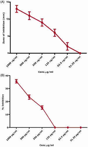
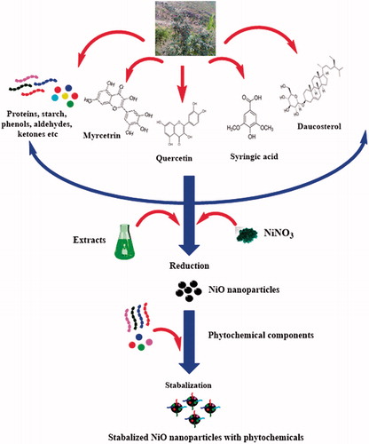
Figure 14. Plausible mechanism for the biosynthesis of NiO nanoparticles via green route.