2,411
Views
23
CrossRef citations to date
0
Altmetric
Reviews

Nanocarrier based approaches for targeting breast cancer stem cells

, , & ORCID Icon
Pages 885-898 | Received 22 Jun 2017, Accepted 07 Aug 2017, Published online: 21 Aug 2017

Figures & data

Figure 1. The role of BCSCs in tumour relapse and metastasis. (A) Conventional chemotherapeutic agents for treatment of breast cancer eliminate bulk tumour cells (non-BCSCs) while sparing tumour initiating, BCSCs and results in tumour relapse; (B) The transition from an epithelial state to a mesenchymal state (EMT) in BCSCs (or metastatic cells) occurs by activation of transcription factors such as Snail, Slug and Zeb and promotes dissemination. This EMT phenotype of cells enters blood circulation and survives by evading immune surveillance, then enter target organ. These cells can remain in a dormant state, lasting from months to decades or re-acquire epithelial properties by undergoing mesenchymal to epithelial transition (MET) in target organ to initiate secondary tumour growth; (C) Key signalling pathways implicated in the survival of BCSCs.

Figure 1. The role of BCSCs in tumour relapse and metastasis. (A) Conventional chemotherapeutic agents for treatment of breast cancer eliminate bulk tumour cells (non-BCSCs) while sparing tumour initiating, BCSCs and results in tumour relapse; (B) The transition from an epithelial state to a mesenchymal state (EMT) in BCSCs (or metastatic cells) occurs by activation of transcription factors such as Snail, Slug and Zeb and promotes dissemination. This EMT phenotype of cells enters blood circulation and survives by evading immune surveillance, then enter target organ. These cells can remain in a dormant state, lasting from months to decades or re-acquire epithelial properties by undergoing mesenchymal to epithelial transition (MET) in target organ to initiate secondary tumour growth; (C) Key signalling pathways implicated in the survival of BCSCs.

Figure 2. Possible therapeutic approaches for eradication of BCSCs using nanocarriers. (1) Targeting BCSC surface markers; (2) Silencing self-renewal pathways; (3) inhibiting drug efflux transporters; (4) inhibiting anti-apoptotic proteins; (5) altering metabolism; (6) targeting autophagy process; (7) disruption of vascular niche of the BCSCs to impair the specialized microenvironment housing.

Figure 2. Possible therapeutic approaches for eradication of BCSCs using nanocarriers. (1) Targeting BCSC surface markers; (2) Silencing self-renewal pathways; (3) inhibiting drug efflux transporters; (4) inhibiting anti-apoptotic proteins; (5) altering metabolism; (6) targeting autophagy process; (7) disruption of vascular niche of the BCSCs to impair the specialized microenvironment housing.

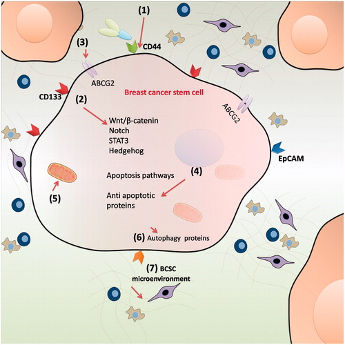
Table 1. Molecular targets of BCSCs.

Table 2. Various anti-BCSCs agents and their mechanism of action for elimination of BCSCs.

Figure 3. The preparation procedure of HA modified mesoporous silica nanoparticles (MSN). MSN modified with an amine-containing silane were first loaded with docetaxel or 8-hydroxy quinoline. Liposomes were prepared with different components consisting of DOPC, DOPE, cholesterol and 18:0 PEG-2000 PE (60:5:30:5 mass ratio). HA was conjugated to the liposomes by an EDC-based method. Finally, hyaluronan modified mesoporous silica nanoparticle-supported lipid bilayers (HA-MSS) was constructed by fusing HA-modified liposomes to drug loaded MSN [Citation31] (Reproduced with permission. Copy Right, Elsevier 2013).

Figure 3. The preparation procedure of HA modified mesoporous silica nanoparticles (MSN). MSN modified with an amine-containing silane were first loaded with docetaxel or 8-hydroxy quinoline. Liposomes were prepared with different components consisting of DOPC, DOPE, cholesterol and 18:0 PEG-2000 PE (60:5:30:5 mass ratio). HA was conjugated to the liposomes by an EDC-based method. Finally, hyaluronan modified mesoporous silica nanoparticle-supported lipid bilayers (HA-MSS) was constructed by fusing HA-modified liposomes to drug loaded MSN [Citation31] (Reproduced with permission. Copy Right, Elsevier 2013).

Table 3. Targeting BCSC surface markers using nanocarriers.

Table 4. Targeting self-renewal pathways of BCSCs using nanocarriers.

Figure 4. Potential nanocarrier systems for efficient targeting of BCSCs. (A) Nanocarriers serve as vehicles for therapeutic agents and increase drug accumulation in tumour tissues via EPR effect. (B) Surface modification of nanocarriers using suitable ligand can reduce off-target effects. (C) Stimuli-responsive nanoparticle systems, which are activated upon exposure to tumour microenvironment may further enhance the accumulation of drugs and also increase cellular internalisation. (D) Nanocarrier systems for co-delivery of dual drugs can improve therapeutic efficacy by simultaneous targeting of both BCSCs and non-BCSCs.

Figure 4. Potential nanocarrier systems for efficient targeting of BCSCs. (A) Nanocarriers serve as vehicles for therapeutic agents and increase drug accumulation in tumour tissues via EPR effect. (B) Surface modification of nanocarriers using suitable ligand can reduce off-target effects. (C) Stimuli-responsive nanoparticle systems, which are activated upon exposure to tumour microenvironment may further enhance the accumulation of drugs and also increase cellular internalisation. (D) Nanocarrier systems for co-delivery of dual drugs can improve therapeutic efficacy by simultaneous targeting of both BCSCs and non-BCSCs.

Reprints and Corporate Permissions

Please note: Selecting permissions does not provide access to the full text of the article, please see our help page How do I view content?

To request a reprint or corporate permissions for this article, please click on the relevant link below:

Academic Permissions

Please note: Selecting permissions does not provide access to the full text of the article, please see our help page How do I view content?

Obtain permissions instantly via Rightslink by clicking on the button below:

If you are unable to obtain permissions via Rightslink, please complete and submit this Permissions form. For more information, please visit our Permissions help page.