
Figures & data
Figure 1. The expression levels of MTAP in colorectal cancer cell lines. Relative expression of MTAP and TLR4 mRNA in colorectal cancer cell lines and normal colonic myofibroblasts cells by Q-PCR. The results indicated that MATP was upregulated in colorectal cancer cell lines. *indicated p < .05.
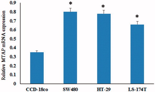
Figure 2. MTAP accelerated tumour cell proliferation and migration. (A) Successful construction of MTAP high expressed and low expressed in SW40 cells; (B) the proliferation ability of MTAP-high-expressed and low-expressed cells were detected by MTT assay; (C and D) the migration and invasive ability of MTAP high-expressed and low-expressed cells were detected. *indicated p < .05.
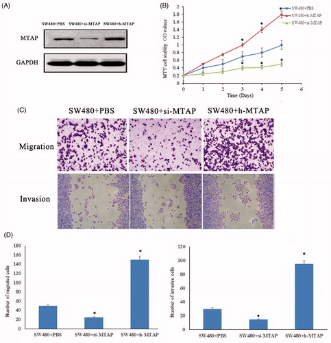
Figure 3. High protein expression of MTAP in colorectal cancer. The expression of MTAP was significantly increased in colorectal cancer tissues compared to paracancerous tissues by IHC. *indicated p < .05.
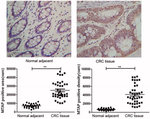
Figure 4. High expression of MTAP accelerated the liver metastasis of colorectal cancer. IHC examinations indicated increased expression levels of MTAP and the mesenchymal markers fibronectin, vimentin and slug and decreased expression of epithelial marker E-cadherin in the clinical liver metastasis specimens compared to the primary tumor (×200). *indicated p < .05.
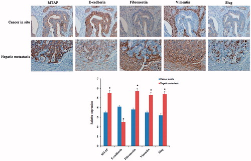
Table 1. Statistics of MTAP in 23 cases of liver metastatic colorectal cancer patients.