8,667
Views
46
CrossRef citations to date
0
Altmetric
Review

Nanomedicine approaches for sirolimus delivery: a review of pharmaceutical properties and preclinical studies

, , , &
Pages 1-14 | Received 17 Aug 2017, Accepted 18 Nov 2017, Published online: 29 Nov 2017

Figures & data

Figure 1. Schematic illustration of the mammalian target of rapamycin (mTOR) pathway and sirolimus (SIR) mechanism. mTOR integrates in/out from numerous upstream cues including several growth factors and interleukin-2 (IL-2). After entry into the cell, sirolimus associates with FK506 binding protein 12 (FKBP-12). The resulting complex interacts with the FKBP12-rapamycin binding domain located in the carboxyl terminus of mTOR and inhibits the mTOR kinase activity.

Figure 1. Schematic illustration of the mammalian target of rapamycin (mTOR) pathway and sirolimus (SIR) mechanism. mTOR integrates in/out from numerous upstream cues including several growth factors and interleukin-2 (IL-2). After entry into the cell, sirolimus associates with FK506 binding protein 12 (FKBP-12). The resulting complex interacts with the FKBP12-rapamycin binding domain located in the carboxyl terminus of mTOR and inhibits the mTOR kinase activity.

Figure 2. Indications for which sirolimus has been investigated. A growing number of evidence has demonstrated that sirolimus is beneficial for treating various diseases [Citation13–27]. GVHD: Graft-versus-host disease; HIV: Human immunodeficiency virus; LAM: Lymphangioleiomyomatosis.

Figure 2. Indications for which sirolimus has been investigated. A growing number of evidence has demonstrated that sirolimus is beneficial for treating various diseases [Citation13–27]. GVHD: Graft-versus-host disease; HIV: Human immunodeficiency virus; LAM: Lymphangioleiomyomatosis.

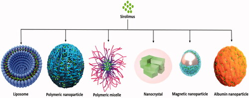
Figure 3. Schematic representation of various nanocarriers for sirolimus delivery which are currently under in vitro and preclinical evaluation.

Figure 3. Schematic representation of various nanocarriers for sirolimus delivery which are currently under in vitro and preclinical evaluation.

Table 1. Nanocarrier compositions and characteristics of sirolimus liposomal formulations.

Figure 4. Chitosan decorated sirolimus liposomes attenuated vascular restenosis following local delivery as confirmed by pathology, immunohistochemical and in vivo CT angiography. Reprinted from ref [Citation46]. Copyright (2017), with permission from Elsevier.

Figure 4. Chitosan decorated sirolimus liposomes attenuated vascular restenosis following local delivery as confirmed by pathology, immunohistochemical and in vivo CT angiography. Reprinted from ref [Citation46]. Copyright (2017), with permission from Elsevier.

Figure 5. Schematic structure of the theranostic targeted thermosensitive liposome (A) and its antitumour mechanism (B). Redrawn with permission from Pang et al. [Citation47]. Copyright (2016) American Chemical Society.

Figure 5. Schematic structure of the theranostic targeted thermosensitive liposome (A) and its antitumour mechanism (B). Redrawn with permission from Pang et al. [Citation47]. Copyright (2016) American Chemical Society.

Table 2. Nanocarrier compositions and characteristics of sirolimus polymeric nanoparticles.

Table 3. Nanocarrier compositions and characteristics of sirolimus micelles.

Table 4. Nanocarrier compositions and characteristics of sirolimus incorporated in miscellaneous nanoformulations.

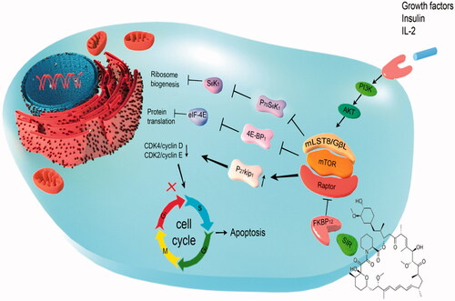
Figure 6. Design of sustained sirolimus delivery nanomedicines based on acetalated β-CD (A), sirolimus concentrations in the blood and the aorta of C57BL/6 mice (n = 4, mean ± SEM), after subcutaneous administration of sirolimus nanoparticles at 3 mg/kg of drug (B). Therapy with sirolimus nanoparticle significantly reduced atherosclerosis in ApoE−/−mice (C). (C) Representative photographs of total aortas from each group and quantitative results subjected to various treatments. Reprinted with minor modification from Dou et al. [Citation97]. Copyright (2016), with permission from Elsevier.

Figure 6. Design of sustained sirolimus delivery nanomedicines based on acetalated β-CD (A), sirolimus concentrations in the blood and the aorta of C57BL/6 mice (n = 4, mean ± SEM), after subcutaneous administration of sirolimus nanoparticles at 3 mg/kg of drug (B). Therapy with sirolimus nanoparticle significantly reduced atherosclerosis in ApoE−/−mice (C). (C) Representative photographs of total aortas from each group and quantitative results subjected to various treatments. Reprinted with minor modification from Dou et al. [Citation97]. Copyright (2016), with permission from Elsevier.

Reprints and Corporate Permissions

Please note: Selecting permissions does not provide access to the full text of the article, please see our help page How do I view content?

To request a reprint or corporate permissions for this article, please click on the relevant link below:

Academic Permissions

Please note: Selecting permissions does not provide access to the full text of the article, please see our help page How do I view content?

Obtain permissions instantly via Rightslink by clicking on the button below:

If you are unable to obtain permissions via Rightslink, please complete and submit this Permissions form. For more information, please visit our Permissions help page.