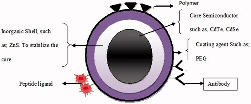
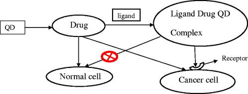
Figures & data
Table 1. Summary of optical properties of different nanosystems in diagnostics [Citation5].
Zhang Z, Hao J, Zhang J, et al. Protein as the source for synthesizing fluorescent carbon dots by a one-pot hydrothermal route. RSC Adv. 2012;2:8599–8601. Chen G, Wu S, Hui L, et al. Assembling carbon quantum dots to a layered carbon for high-density supercapacitor electrodes. Scientific Rep. 2016;6:19028. Sun YP, Wang X, Lu F, et al. Doped carbon nanoparticles as a new platform for highly photoluminescent dots. J Phys Chem C. 2008;112:18295–18298. Yang ST, Wang X, Wang H, et al. Carbon dots as nontoxic and high-performance fluorescence imaging agents. J Phys Chem C. 2009;113:18110–18114. Wang X, Qu K, Xu B, et al. Microwave assisted one-step green synthesis of cell-permeable multicolor photoluminescent carbon dots without surface passivation reagents. J Mater Chem. 2011; 21:2445–2450. Sun Y-P, Wang X, Lu F, et al. Doped carbon nanoparticles as a new platform for highly photoluminescent dots. J Phys Chem C, Nanomater Interfaces. 2008;112:18295–18298. Ray SC, Saha A, Jana NR, et al. Fluorescent carbon nanoparticles: synthesis, characterization, and bioimaging application. J Phys Chem C. 2009;113:18546–18551. Parak WJ, Boudreau R, Le Gros M, et al. Cell motility and metastatic potential studies based on quantum dot imaging of phagokinetic tracks. Adv Mater. 2002;14:882–885. Michalet X, Pinaud FF, Bentolila LA, et al. Quantum dots for live cells, in vivo imaging, and diagnostics. Science. 2005;307:538