Figures & data
Figure 1. SEM morphologies of MAO (a, b) and MHTZn coating (c, d).
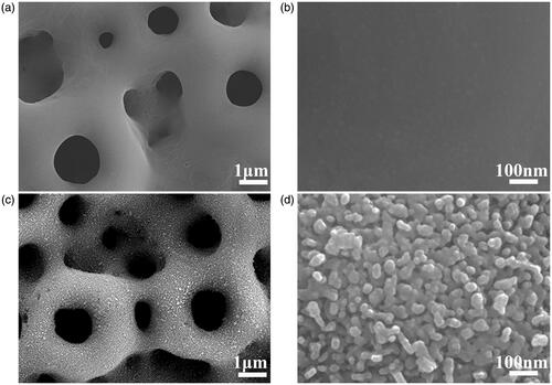
Figure 2. Morphologies of attached RAW264.7 cells cultured on MAO coating for one day (a), and three days (b), and on MHTZn coating for one day (c) and three days (d).
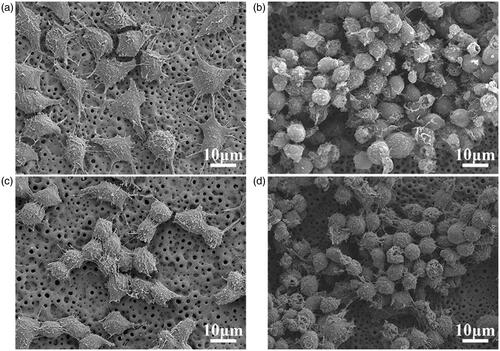
Figure 3. Skeleton and nuclei staining for MAO group at day 1 (a), day 3 (b), and day 5 (c); MHTZn group at day 1 (d), day 3 (e), and day 5 (f). Cell viability of RAW264.7 cells on MAO and MHTZn coatings tested by CCK-8 (g). Values are mean ± SD (n=3); * depicts statistical differences; * p < .05 vs. MAO.
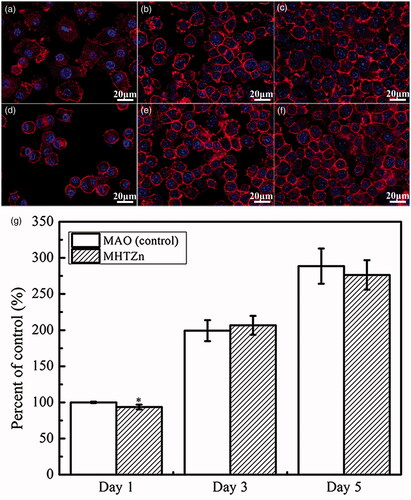
Figure 4. Inflammatory secretions of RAW264.7 cells cultured on MAO and MHTZn coatings for two days. Values are mean ± SD (n = 3); *statistical differences; *p < .05, **p < .01 vs. MAO.
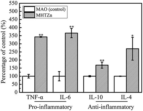
Figure 5. SaOS-2 cells after culturing in the MAO conditioned media for one day (a) and three days (c), in the MHTZn conditioned media for one day (b) and three days (d), showing the nuclei stained by DAPI and the cytoskeleton stained by phalloidin with TRITC.
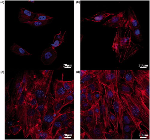
Figure 6. Cell viability of SaOS-2 cells cultured in the MAO and MHTZn conditioned media tested by CCK-8. Values are mean ± SD (n = 3).
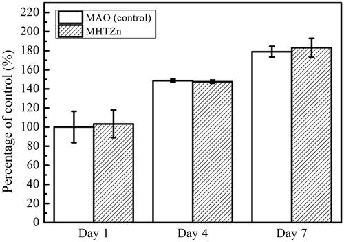
Figure 7. ALP activity of SaOS-2 cells cultured in MAO and MHTZn conditioned media for 7 days. MAO group used as the control (a), MHTZn group (b) and OD values (c). Values are mean ± SD (n=3); depicts statistical differences; * p < .05 vs. MAO.
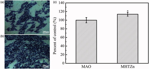
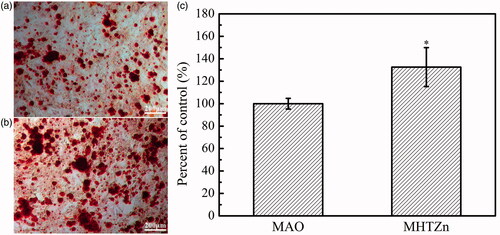
Figure 8. Extracellular mineralization of SaOS-2 cells cultured in the MAO and MHTZn conditioned media for 14 days. MAO group used as the control (a), MHTZn group (b) and OD values (c). Values are mean ± SD (n = 3); * depicts statistical differences; * p < 0.05 vs. MAO.