Figures & data
Figure 1. Characterization of functional docetaxel nanomicelles. (A) Structural representation of functional docetaxel nanomicelles; (B) size distribution; (C) atomic force microscope image; (D) transmission electron microscope image; (E) release rates of docetaxel from nanomicelles. Data are presented as mean ± standard deviation (SD) (n = 3).
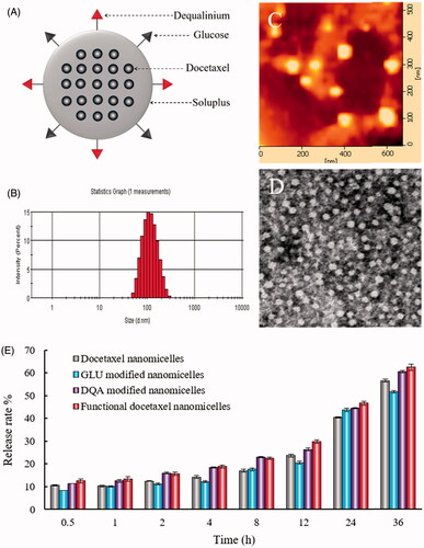
Table 1. Characterization of the functional docetaxel nanomicelles.
Figure 2. Transport ability of functional docetaxel nanomicelles across the BBB model. (A) Schematic representation of the BBB model. (B) Functional docetaxel nanomicelles transporting across the BBB. (C) Transport ability indicated by the survival rate of brain glioma U87MG cells after drug transport across the BBB. p < .05, a, vs. 1; b, vs. 2; c, vs. 3. Data presented as mean ± standard deviation (n = 3). BBB is the abbreviation of the blood–brain barrier; BMVECs is the abbreviation of the brain microvascular endothelial cells; DQA is dequalinium.
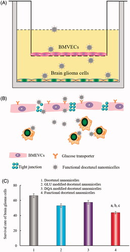
Figure 3. Cellular uptake by brain glioma cells. (A) Cellular uptake by brain glioma U87MG cells observed with flow cytometry. (a) Coumarin nanomicelles; (b) GLU modified coumarin nanomicelles; (c) DQA modified coumarin nanomicelles; (d) functional coumarin nanomicelles. (B) Cellular uptake by brain glioma U87MG cells under fluorescence microscope. (B1) Coumarin nanomicelles; (B2) GLU modified coumarin nanomicelles; (B3) DQA modified coumarin nanomicelles; (B4) Functional coumarin nanomicelles. Courmarin was used as a fluorescent probe.
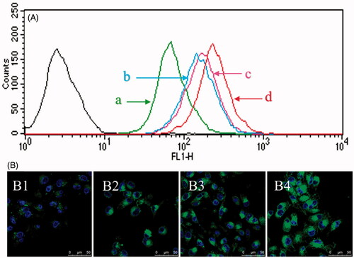
Figure 4. Cytotoxicity to brain glioma cells and the glioma spheroids after treatment with functional docetaxel nanomicelles. (A) Cytotoxic effect to brain glioma U87MG cells; p < .05, a, vs. docetaxel nanomicelles; b, vs. GLU modified docetaxel nanomicelles; c, vs. DQA modified docetaxel nanomicelles. Data presented as mean ± standard deviation (SD), (n = 6). (B) Cytotoxic effect to brain glioma U87MG glioma spheroids. (B1) Docetaxel nanomicelles; (B2) GLU modified docetaxel nanomicelles; (B3) DQA modified docetaxel nanomicelles; (B4) functional docetaxel nanomicelles.
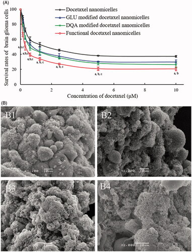
Figure 5. Induction of apoptosis on brain glioma cells after treatment with functional docetaxel nanomicelles. (A) Induction of apoptosis; (B) activation of apoptotic enzymes upstream caspases 8, 9 and downstream caspase 3. 1. Blank control; 2. Docetaxel nanomicelles; 3. GLU modified docetaxel nanomicelles; 4. DQA modified docetaxel nanomicelles; 5. Functional docetaxel nanomicelles. p < .05, a, vs. 1; b, vs. 2; c, vs. 3; d, vs. 4. Data presented as mean ± standard deviation (SD), (n = 3).
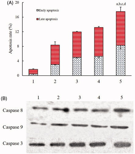
Figure 6. Co-localization of functional coumarin nanomicelles with mitochondria of brain glioma cells. (A) Co-localization with mitochondria of brain glioma U87MG cells; (B) Quantitative analysis of co-localization rates.
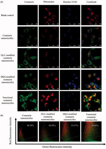
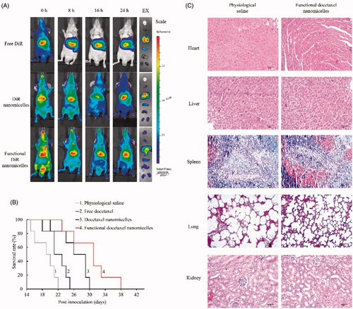
Figure 7. In vivo imaging and efficacy on brain glioma-bearing nude mice after treatment with functional nanomicelles. (A) In vivo imaging of glioma-bearing nude mice after administration of functional DiR nanomicelles; (B) Kaplan–Meier survival curves of brain glioma-bearing nude mice after treatment with functional docetaxel nanomicelles; (C) Preliminary safety evaluation on major organs after treatment with physiological saline and functional docetaxel nanomicelles (hematoxylin-eosin staining).