Figures & data
Figure 1. Schematic diagram of electrostatic spinning of the composite polypropylene mesh.
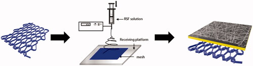
Table 1. The mechanical strength and parameters of the PP mesh.
Figure 2. Mechanical properties and construction of the mesh. (A) Effects of different concentrations of RSF solution on electrospinning. (B,C) The pore size was controlled in a limited area. (D–F) The mechanical properties of the two types of mesh. No significant differences were observed between RSF and PCL fibres.
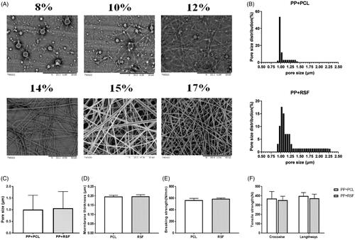
Figure 3. (A,B) PP mesh with electrostatic spinning did not increase cytotoxicity. (C) SEM shows that the L6 cells adhered better on the RSF fibre across the area of the cell than on the other surfaces.
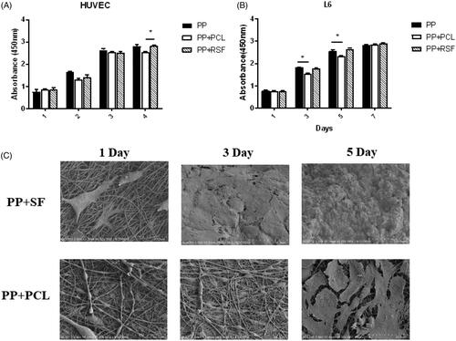
Figure 4. (A) The result of tissue repair in a full-thickness abdominal wall defect model. (B,C) The area and score of mesh adhesion. The RSF fibre could significantly reduce adhesion.
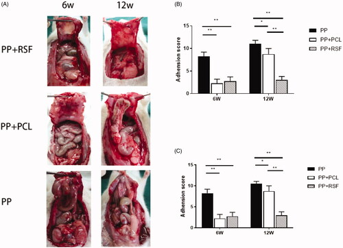
Figure 5. The RSF electrospun membrane can efficiently regenerate the abdominal wall. The black arrow shows the mesh covering a complete tissue overlay. The red arrow shows adhesion of the bowel around the mesh.
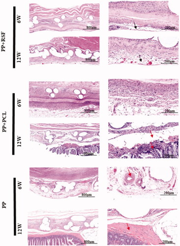
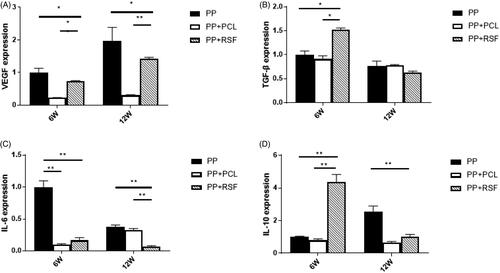
Figure 6. The RNA expression results. RSF exhibits low expression of the peritoneal inflammatory factor IL-6 and promotes the expression of VEGF, TGF-β, and IL-10.