
Figures & data
Scheme 1. Synthesis of poly-(xylitol-g-adipate-co-glutamide) (PXAG) copolymer.
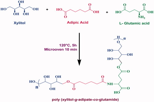
Figure 1. FTIR spectrum of (a) PXAG, (b) PXAG-PHB and (c) PXAG-PHB/FFE scaffold.
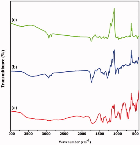
Figure 2. The 1H (proton) NMR spectrum of the PXAG copolymer.
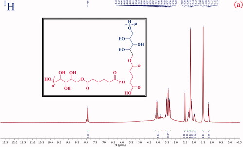
Figure 3. The carbon (13C) NMR spectrum of the PXAG copolymer.
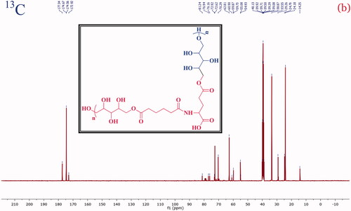
Figure 4. XRD spectra of (a) PXAG, (b) PXAG-PHB and (c) PXAG-PHB/FFE polymers.
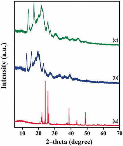
Figure 5. SEM images of the PXAG (a) PXAG-PHB scaffold (b, e) and PXAG-PHB/FFE (c, f) via ultra-sonication method and magnetic stirrer method, respectively. The image: d is the magnified observation of the PXAG-PHB scaffold by the ultra-sonication processes.
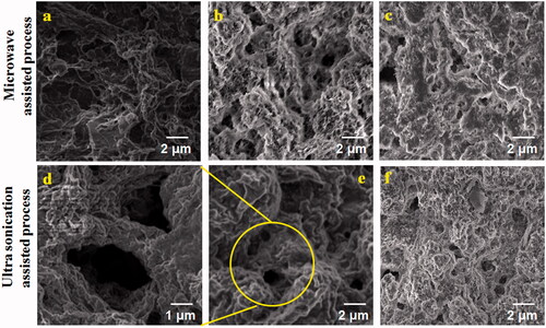
Figure 6. The cell viability of PXAG, PXAG-PHB and PXAG-PHB/FFE scaffold in normal wound cells at 100 μg/mL.
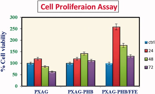
Figure 7. Nuclear assessment by Hoechst 33258 staining in diabetic wound cell model preserved with 100 μg/mL of PXAG, PXAG-PHB and PXAG-PHB/FFE scaffold.
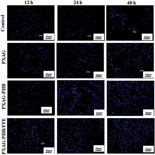
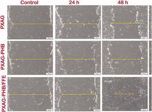
Figure 8. Scratch assay images of 100 μg/mL of PXAG, PXAG-PHB and PXAG-PHB/FFE scaffold treated diabetic wounded cell models using different incubation time intervals (control, 24, 48 and 72 h).
Supplemental Material
Download MS Word (2.8 MB)Data availability statement
The authors confirm that the data supporting this study's findings are available within the article and its supplementary materials.